Download
1 / 23
230 likes | 289 Vues
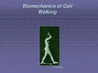
Walking Gait Cycle. Walking Gait Cycle - 60:40 stance to swing phase Stance Phase: (IC) LR – MS – TST – PSW LR – beginning of 1st double support phase MS – foot is in full contact – adapting to env’t, beginning of single support, which is of equal

E N D
Walking Gait Cycle • Walking Gait Cycle - 60:40 stance to swing phase Stance Phase: (IC) LR – MS – TST – PSW LR – beginning of 1st double support phase MS – foot is in full contact – adapting to env’t, beginning of single support, which is of equal duration of contralateral swing phase TST – foot is preparing to toe off (TO) PSW – 2nd double support phase Swing Phase: begins with TO and ends w/ IC ISW – MSW - TSW
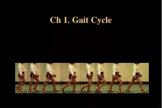
Running vs. Walking Gait Cycles • The Running Gait Cycle has a temporal reversal of Stance:Swing phases (40:60) as compared to Walking Gait Cycle (60:40); the stance phase during sprinting may be as low as 22% of cycle Stance Phase: Absorption – (Mid stance) – Propulsion Swing Phase: ISW (75%) – (MSW) - TSW (25%) Running Gait – two periods of double float in swing; refers to when neither foot is in contact w/ the ground; at the beginning and at the end of each running swing phase Walking Gait – two double support periods in stance
Running Gait Cycle • Step length – IC of one foot to IC of the 2nd foot • Stride length – IC of 1st foot to IC of the same foot • Cadence – number of steps in a given time; on average about 100-122 steps/min with females averaging about 6-9 s/m higher • As running Velocity increases, there is an initial increase in step length, followed by increased cadence • Stride length is limited by runner’s leg length, height, and ability; generally the longer the stride, the higher the velocity • When optimum stride length is attained; further velocity increases will come from increased cadence
Kinematics • Kinematics of Walking and Running are much different • There is an increase in joint ROM with increasing velocity • Virtually no difference is found in the transverse and frontal plane kinematics; with most of the difference occurring in the sagittal plane • Lower C of G • Increased speed due to increased flexion of hips and knees; and increased dorsiflexion of the ankle
Knee Kinematics of Running • The knee demonstrates increased flexion with increasing velocity, but as seen with the hip, extension decreases • Absorption phase of the stance phase sees knee flexion to accommodate ground reactive forces; walking only requires about 10 deg of flexion vs.35 during running • Max knee flexion occurs at MS, after IC, during the absorption phase; this is followed sequentially by knee ext; max knee flexion during walking occurs just after TO • Avg. Knee ROM is 63 deg during Running and 60 deg during walking; the major difference is that max flexion during walking only reaches an avg. of 64 deg, whereas during running it reaches an avg. of 79 deg.; conversely, knee extension is on average, 10 degrees less during running than during walking (-16 deg. vs -6 deg).
Hip Kinematics of Running • Flexion of the hip increases, as extension of the hip actually decreases with increasing velocity • One study of walking found overall ROM of 43 deg, with 37 deg of flexion and 6 deg of ext; this study also found an increased ROM during running, with overall ROM averaging 46 deg, all of which was hip flexion with the hip never reaching neutral (negative extension) • Max hip ext occurs at TO; Max hip flex occurs at TSW
Ankle and Foot Kinematics • Ankle joint – primary plantar/dorsiflexor • Foot joints – including subtalar, oblique midtarsal, longitudinal midtarsal and 5th ray; provide for tri-planar pronation/supination • Pronation – dorsiflexion/eversion/abduction • Supination – plantarflexion/inversion/adduction • Metatarsalphalangeal joints (MTP) are biplanar – mostly dorsiflexion/plantarflexion w/ some abd/add
Ankle and Foot Kinematics cont. • Walking: ankle plantarflexes after IC and during LR, followed by dorsiflexion at MS; overall ROM is approx. 30 deg (18 plantarflex/12dorsiflex) • Running: overall ankle ROM of 50 deg; • At IC (rearfoot in most), ankle undergoes rapid dorsiflexion during absorption (pronation) • Supination is limited due to diminished time of plantarflexion, and pronation is increased • May lead to excessive pronation injuries • Running shoes or orthotics may limit this excessive pronation, and allow for more supination, and thus a more rigid foot for propulsion • A pronated subtalar joint allows the foot to become the “mobile adapter”; whereas a supinated subtalar joint serves to lock the midtarsal joints, creating a rigid lever to better serve propulsion
Windlass Mechanism The plantar fascia and the intrinsic foot muscles increase the efficiency of propulsion by providing “spring-like” support to the medial arch of the foot, helping to deliver the foot into supination, and contributing an elastic tension.
Lower Extremity Kinematics of Running • At IC, the pelvis, femur and tibia begin to internally rotate; int. rotation lasts through LR until MS; this everts and unlocks the subtalar joint, oblique and longitudinal midtarsal joints and in turn absorbs shock (pronation) • External Rotation of the pelvis, femur and tibia begin following MS, causing inversion and subtalar and mid foot locking, creating the rigid lever for propulsion • All lower extremity joints work together during walking/running to provide a biomechanically efficient means of locomotion • These joints depend on each other and upon muscular action to carry out walking or running
Lower Extremity Kinematics of Running (cont.) Metatarsal Break- the oblique line drawn across the metatarsal heads. This oblique axis promotes hind foot inversion during toe off, which contributes to external rotation of the entire stance leg
Lower Extremity Kinetics • Kinetics – the study of forces that cause movement, both internally (muscular) and externally (ground reactive forces) • As compared to walking, running increases muscle activity in all muscles • Ground reactive forces – measured with a force plate system – demonstrates that vertical reactive forces are the most significant in running • In rearfoot or heel strikers (80% of runners), there is a “two-bump” force plate appearance with one occurring in the rearfoot during loading response and one in the forefoot during propulsion • Walking produces GRF of 1.3-1.5x body weight • Running produces GRF of 3-4x body weight
Clinical Note • Running injuries typically occur as a result of volume training • With 3-4x body weight with each impact, 50-70 steps per foot per minute, 300-900 times per mile, the cumulative load can be measured in tons • Stress fractures occur as a result of high volume training, combined with inadequate rest and recovery and/or biomechanical flaws • Observed running injuries often occur at sites that mirror areas of peak force plate measures • Placing a runner in a cushioning shoe may minimize peak force; however, the extra shock absorbing materials built into the midsole may result in excessive pronation in some runners, both due to less restriction of pronation and possibly due to increased moment arm on which GRFs act
Running Economy • Measured in terms of Submaximal Metabolic Energy Expenditure (VO2submax), running economy is a method by which running biomechanics are studied to determine their affect on running performance • It is hypothesized that some variations in economy might be due to differences in genetic factors that cannot be changed through technique adjustments or training • Other factors that are thought to contribute include motor unit recruitment, anatomical mechanical advantage and movement skill
Factors and their Positive Effect on Running Economy Vertical oscillation Pronation Trunk lean A-P Impulse Plantarflexion Knee Extension Plantarflexion velocity Arm motions Vertical Force Hip Extension Stride Length Stride Index HIGH VO2 LOW
Running Economy (cont.) • It is likely that Running Economy is directly effected by running mechanics • However, it is not known how much running performance can be enhanced by altering a runner’s technique or style • Many running related movement patterns that may seem uneconomical or sub-optimal may be as a result of an adaptation to a structural or functional anomaly; where alteration of that pattern may diminish economy and/or increase risk of injury • External factors that can influence economy include shoe weight, midsole composition, wind velocity, materials and slope of running surface. These have been identified and are relatively easy to measure. • Identifying running styles or techniques that can predictably result in economy changes is very difficult
References • O’Connor, F and Wilder, R (2001). Textbook of Running Medicine, McGraw Hill. • Neumann, D.A. (2002). Kinesiology of the Musculoskeletal System. St. Louis, Missouri. Mosby. • McGinnis, P.M. (2005). Biomechanics of Sport and Exercise 2nd ed. Champaign, IL. Human Kinetics. • Cavanagh, P.R. (1990). Biomechanics of Distance Running. Champaign, IL. Human Kinetics