Hemoglobin
Hemoglobin. DR QAZI IMTIAZ RASOOL. OBJECTIVES Describe the structure of hemoglobin molecule Describe synthesis of hemoglobin in brief. List the functions of hemoglobin. Enlist various types of normal and abnormal hemoglobin’s. Hemoglobin. 1. if the body had to depend upon


Hemoglobin
E N D
Presentation Transcript
Hemoglobin DR QAZI IMTIAZ RASOOL
OBJECTIVES • Describe the structure of hemoglobin molecule • Describe synthesis of hemoglobin in brief. • List the functions of hemoglobin. • Enlist various types of normal and abnormal hemoglobin’s.
Hemoglobin 1. if the body had to depend upon dissolved O2 in the plasma to supply O2 to the cells 2.The heart would have to pump 140 l/min instead of 4-6 l/min. 3. RBC have nuclei during early stages of development mature to make room for Hb
HB historical facts • 1stprotein to be crystallized (1849); • 1stto be associated with a specific physiological function (1875); • 1stproteins to have its molecular weight (64,500) determined correctly (1920s); • 2nd protein having its 3-D structure determined (1969) • .Hb: A protein that you can “see” with your naked eyes!
Synthesis: following R essential 1.1st class proteins – milk, fish, egg, soyabean etc. 2. Metals – iron and sulphur.------copper+calcium 3. Vitamin B12 (Folic acid).-------cobalt 4. Porphyrin compound
Synthesis 1.- Erythroblasts, =65% ---7days—intermediate normoblast stage - Reticulocyte stage=35% ------2 days 2. Haem synthesis in the mitochondria. 3. Globin synthesis in the polyribosomes 4. Although synthesis occur separately within developing red cell precursors, their rates R coordinated for Hb assembly.
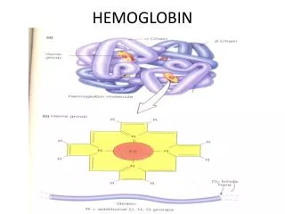
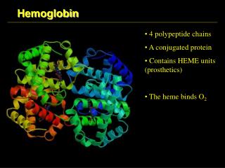
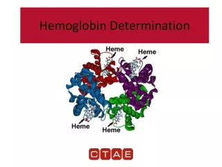
HaemoglobinChemistry: It is a chromo protein, mol:wt. 64000 1.Single RBC contains about 270 million HB molecules. Each RBC can hold about 1 billion mol of O2. Hb =1/3 of the weight of RBC 2. Heme = porphyrin ring + Fe2 non-protein (pigment) • Source of iron in body • Iron – held in organic lattice • Each iron holds 1 mol O2 3. Adult HbGlobin= 2 α+ 2β chains each with its own haem group i.e. α1 β1and α2 β2
Structure of Heme 1.a protoporphyrin ring with an iron atom at its centre starts from a cyclic tetrapyrrole i.e. consists of 4 mol; of pyrrole (mol:wt1600 X 4). 2. - imparts a red color 3. -R, -methyl (M), -vinyl (V) - proprionate (Pr) groups Pyrrole ring
Iron • Food: Fe3+ absorbable Fe2+ `Gastric juice (gastroferrin)+ vit C reduces • Absorbed in duodenum • Fe2+plasma level 10-35 mol/l • Apoferritin (mucosa) • Transferrin ( Fe3+; plasma; β1-globulin), • Ferritin; spleen, liver, bone marrow; plasmaferritin, • hemosiderin • There is no physiological mechanism for the excretionof excess iron
Iron metabolism • Iron is indipensable for life (either in heme or non-heme form essential for oxygen transport, electron transfer, DNA synthesis, etc.) 2.Iron is insoluble ([Fe] cannot exceed 10-17) 3.Iron is potentially toxic (unless appropriately chelated, Fe plays a key role in the formation of oxygen radicals)
GENETIC ROLE Humans normally carry 8 functional globin genes, arranged in 2 duplicate gene clusters: These genes code for 6 different types of globin chains: α,β,γ,δ,ε,ζ, . 2 α (each with141 amino acids) 6. 2 β each - 146
Normal Hb in adult blood HbAIC: glycated Hb – important marker of long-term diabetes compensation
Haemoglobin catabolism • Destruction - life span- 120 days. • removed extravascularly by macrophages of the reticuloendothelial system (RES), - specially in the bone marrow - but also in the liver and spleen. 3. cell metabolism deteriorates as enzymes R degraded and not replaced, makes it non viable, but the exact reason why the red cells die is obscure.
HEMOGLOBIN BREAKDOWN MACROPHAGE
HB breakdown haemoglobin haem globin iron protoporphyrin Amino acids Bilirubin (free) CO Expired air transferrin Liver conjugation erythroblast Bilirubin glucuronides Enterohepatic cir Urobilin(ogen) Stercobilin(ogen) Urine faeces
FUNCTIONS 1. - combines reversibly with O2to form oxy-HB (HbO2) . 2. combines + CO2 and transports 30% of total as(carbamino-HB) 3. Acts as a buffer 4. forms typical haem crystals with NaCl and glacial acetic acid which is useful in diagnosis of blood sample (man or animal). 5. combines with other gases like CO, H2S ( poisoning ,death) 6.Nitricoxide(NO) binds to Hb, NO causing vasodilation to ↑ blood flow and O2 delivery
Normal values • At birth: 23 gm% • Falls to 10.5gm% by 3 month (breast feed no iron) • Rises gradually to 12.5 rises at 1 year of age. • Males: 14 – 18 gm% • Females: 12 – 15 gm % females: 12 – 15 gm%
Differences Hemoglobin & Myoglobin • Found in Blood • Composed of 4 Heme and 4 Globin chains • Carrier of Oxygen and Carbon dioxide • Higher Oxygen affinity • Found in Heart and skeletal muscles • Composed of 1 Heme and 1 Globin chain • Reservoir and Carrier of Oxygen • Lesser Oxygen affinity
Glu6Val6 oxyMb (MbO2) oxyHb (HbO2) deoxyMb deoxyHb O2 O2 O2 O2 O2 Glu6Val6 Myoglobin and HbStructure
CLASSIFICATION of ANEMIASDeficiency of Hb in the blood caused by either: RBC Count or Hb in the RBCs • Hemorrhagic anemiahemorrhagic anemia • Aplasticanemiaaplastic anemia • Megaloblasticanemiamegaloblastic anemia • Pernicious anemiapernicious anemia • Hemolytic anemiahemolytic anemia • Sickle cell anemiasickle cell anemia • Iron deficiency anemiairon deficiency anemia • Secondary anemia (renal)secondary anemia (renal)
Anemia – reduced O2 carrying capacity of the blood • Insufficient hemoglobin content in RBCs: Iron Deficiency - inadequate intake or absorption of iron. Pernicious - dietary deficiency of Vitamin B12 or inadequate production of intrinsic factor for absorption of Vitamin B12.
Abnormal hemoglobin • Sickle Cell - one amino acid in the 287 forming the beta chains is wrong . In low O2 conditions the beta chains form stiff rods which cause RBCs to sickle blocking small vessels.
Hb S CAUSE: • This disease is caused by a mutation in Hb. (Abnormal polypeptides due to substitution of amino acids) • Supression of synthesis of polypeptide chains. • Occurs in 0.3 to 1 % of west african & american black people • Valine is substituted for glutamic acid at 6 position of beta chain.
Red blood cells from sickle cell anemia patients: become sickle-shaped only in the deoxygenated state!
Thalassemia • Defect in the synthesis of either α & β • If the body doesn't produce enough of either proteins, the RBC cannot carry sufficient O2. • - is anemia in early childhood and lasts throughout life.
Porphyria • abnormalities in the chemical steps production of heme • It is characterized by extreme sensitivity to light (expo- sure to sunlight causes vesicular erythema), -reddish-brown urine, - reddish-brown teeth, and ulcers which destroy cartilage and bone, 3.causing the deformation of the nose, ears, and fingers. Mental aberrations such as hysteria, manic-depressive psychosis, and delirium,
Hemoglobine derivates unable to transport CO2 • Methemoglobin: contains Fe 3 instead of Fe2 (e.g. nitrate/nitrite containing food or water) • Carboxyhemoglobin– CO poisoning, smokers (cherry red colour) • Sulfhemoglobin – green