Introduction to Mycology
530 likes | 1.17k Vues
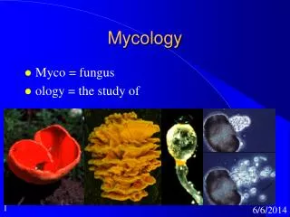
Introduction to Mycology. General Properties of fungi. Fungi are a diverse group of saprophytic and parasitic eukaryotic organisms T hey varies in complexity and size ranging from the single-cell microscopic yeasts to multicellular molds.

Introduction to Mycology
E N D
Presentation Transcript
General Properties of fungi • Fungi are a diverse group of saprophytic and parasitic eukaryotic organisms • They varies in complexity and size • ranging from the single-cell microscopic yeasts to multicellular molds
Fungi can be distinguished from other infectious organisms by • 1- They are Eukaryotes • that is, they have a membrane-enclosed nucleus, and other membrane bounded organelles
2- Cell wall and membrane components • Fungal cell wall are composed largely of Chitin • a polymer of N-Acetylglucosamine, rather than peptidoglycan • The fungal membrane contains ergosterol, rather than the Cholesterol found in mammalian membranes
3- All fungi are Heterotroph, and Chemotrophic organisms • Fungi secrete degradative enzymes ( cellulases, proteases, nucleases) into their immediate environment • 4- Fungi are Facultative anaerobic or obligatory aerobic organisms • 5-Most fungi are Acidophilic organisms • 6- Fungi reproduce and spread through the environment by Spore formation which may be sexual or asexual
Fungi Morphology • Fungi exist into two main forms yeasts or hyphae (Moulds) • Some fungi may occur in both the yeast and mycelialforms • These are called dimorphic fungi • Hyphae are multicellular filamentous structures, constituted by tubular cells with cell walls
Yeast • Yeasts are Unicellular non-branched, oval or rounded cells • measuring 3- 15 µm in diameter Yeast of the species Saccharomyces cerevisiae
Yeasts reproduce asexually by budding (Blastospore, and Chlamydospore) Chlamydospores of the yeastCandida albicans
Dimorphic fungi • These fungi are changing their morphology from mould to yeast phase, or from yeast to mould depending on the growth conditions • 1. Yeast (parasitic or pathogenic form) • This is the form usually seen in tissue, in exudates, or if cultured in an incubator at 37ºC • 2. Mycelium ( saprophytic or mold form) • The form observed in nature or when cultured at 25ºC
Fungal Diseases • Fungal diseases can be classified into the following groups • 1-Hypersensitivity (allergy) • It is an allergic reaction to molds and spores ( Indoor air pollution) • 2-Mycotoxicoses • Poisoning of man and animals due to accidental ingestion of food contaminated by toxic compounds produced by fungi • Examples • A- Ergotism: caused by Clavicepspurpurea; (Ergot alkaloids) • B- Aflatoxicosis: caused by Aspergillus flavus(Aflatoxins)
Ergotism Ergotism
3-Mycetismus • Due to ingestion of Amanitins (a toxin produced by a specific type of mushroom ; Amanita verna) • 4-Infection (Mycosis) • A-Superficial (Hair, skin, nail, cornea) mycosis • B-Subcutaneous mycosis • C-True systemic (endemic) mycosis • D-Opportunistic mycosis
SUPERFICIAL MYCOSIS • The superficial mycoses are usually limited to the outermost layers of the skin, hair, and nails, and do not invade living tissues • Most common Types: • 1- Dermatophytosis • 2- Pityriasisversicolor
Dermatophytosis (Tinea or Ringworm) • It is a type of superficial skin infection of cutaneous layer (mainly epidermis) • Causes: • Epidermophyton • Microsporum • Trichophyton
PityriasisVersicolor • It is a superficial chronic infection of stratum corneum. • This fungal infection is caused by Malassezia furfur
Other superficial infection of skin • TineaNigra, Black Piedra, and White Piedra are caused by • Exophialawerneckii, Piedraiahortae, and Trichosporonspecies respectively Black Piedra TineaNigra White Piedra
1-Tinea Nigra • Exophialawerneckii • Infection of Stratum corneum • 2-Black Piedra • Piedraiahortae • Infections of scalp hair • 3-White Piedra • Trichosporonbeigelii • Fungal infection of facial,axillaryor genital hair
Subcutaneous Mycosis • Sporotrichosis • Caused bySporothrixschenkii • At 25°C: Septatehyphae, rosette-like clusters of conidia at the tips of the hyphae • At 35 C: Yeast
Systemic Mycosis • Respiratory system infection • Chronic granulomatous pneumonia • Causes • Histoplasmacapsulatum(dimorphic fungi) • Coccidioidesimmitis(dimorphic fungi)
Opportunistic mycosis • Candidiasis • Candida albicansinfection • Example: • 1-Oral Candidiasis • 2-Vaginal Candidiasis
Laboratory Diagnosis of Fungal Infections • (1) Specimens • According to the site of infection • Skin scales • Hairs • Nails • Respiratory secretions • Blood
(2) Direct Detection • A- Direct microscopy of unstained preparations (mounting method) • Examination of unstained preparations to demonstrate hyphae, spores or yeast cells • Skin scraping, nails or hairs are mounted with 10-20% KOH • to digest the keratin layer so that hyphae and spores can be seen
B- Direct microscopy of stained preparations • Different stains are used • India ink • Periodic acid-Schiff stain (PAS) • Silver stain • Lactophenol cotton blue stain (specific fungal stain)
(3) Culture • Sabouraud`sdextrose agar (SDA) • The standard media for most fungi • Chloramphenicol added to inhibit bacterial growth & Cycloheximide added to inhibit saprophytic fungi • Incubation temperature is 22°C • If systemic mycosis is suspected, Enriched media is used and incubated at 37°C
(4) Serodiagnosis • Detection of specific antibody help in diagnosis of systemic mycosis • (5) Cutaneous delayed type hypersensitivity test • Example • Histoplasminskin test • Histoplasmacapsulatum • Blastomycinskin test • Blastomycesdermatitidis • For identification of systemic mycosis