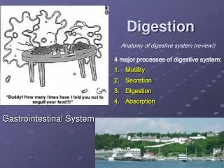
GASTROINTESTINAL FUNCTION
GASTROINTESTINAL FUNCTION. THE STOMACH. A) Helicobacterpylori infection is one of the major cause of peptic ulcer, the ulcer results from weakening of the protective mucous coating of the stomach and duodenum.

GASTROINTESTINAL FUNCTION
E N D
Presentation Transcript
A) Helicobacterpylori infection is one of the major cause of peptic ulcer, the ulcer results from weakening of the protective mucous coating of the stomach and duodenum. • The organism protects itself from gastric acidity by secretion of the urease enzyme which convert the acidic media into more neutral environment.
Tests for H. pylori infection: • 1- Urea breath test: is a non-invasive and reliable test that depends on the urease activity of the organism to detect active Infection. • The patient is given by mouth either 13C- or 14C-labeIled urea and urease (produced by H. pylori) if present, hydrolyses urea into ammonia and isotopically labeled carbon dioxide. • Carbon dioxide is absorbed from the gut and subsequently expired in the breath where it can be detected and quantified.. • This test is used both for theidentificationofpatients with active infection and for establishingtheeffectivenessof treatment.
2- Serological tests: Patients infected with H. pylori develop antibodies to the organism that can be detected in the laboratory using enzyme-linked immunosorbent assays (ELlSAs). • These serological tests are less reliable in confirming the eradication of the organism because of the slow reduction in antibody titers. • Faecal antigen testing: Enzyme immunoassays can be used to detect the presence of H. pylori in stool specimens.
B) Gastrin production: • Gastrin is a polypeptide released by gastric antrum and duodenum and is a potent stimulator of gastric acid production. • Its release is inhibited by low gastric pH , and its circulating levels are increased in patients with chronic hypochlorhydria. • So, plasma gastrin may be elevated in cases of decreased HCL production in the stomach such as due to gastritis, treatment with H2 antagonists, proton pump inhibitors, pernicious anemia or previous vagotomy.
The most important clinical application for the measurement of gastrin is in the investigation of patients with gastric acid hyper secretion thought to be caused by a gastrinoma (Zollinger-Ellison syndrome).
Zollinger-Ellison syndrome • This syndrome is due to a gastrinoma, that is, neoplasia may be due to either: • a) pancreatic gastrin-producing cells (common) • b) gastric gastrin-producing cells • About 60% of gastrinomas are malignant and 30% occur as part of the MEN syndrome type 1. • Increased gastrin production leads to chronic hypersecretion of gastric acid, which in turn causes peptic ulceration and sometimes diarrhea and fat malabsorption leading to steatorrhoea.
The steatorrhoea is believed to be due to inhibition of pancreatic lipase by the high [H+} concentration in the intestinal lumen. • In some patients simple duodenal ulcer or diarrhea only may be the presenting feature.
The diagnosis of gastrinoma is based on the detection of elevated fasting plasma gastrin in association with high gastric acid secretion. • Patients should not be receiving proton pump inhibitors or H2 receptor blocker at the time of measurement. • Provocative testing may be necessary in about 15% of patients where the basal plasma gastrin concentration is normal or only slightly increased and gastrinoma is suspected. • In this provocative test secretin is injected IV, which usually produces a 2-fold increase in plasma gastrin in patients with gastrinoma. while no change occurs in patients with G-cell hyperplasia
Many factors can result in acute pancreatitis but most commonly gallstones or alcoholism is the major ones; vascular and Infective causes have also been recognized. • Plasma amylase activity measurements is the test most commonly used for diagnosis of acute pancreatitis. • Plasma calcium may fall considerably in severe cases of acute pancreatitis, but sometimes not for a few days; it probably falls as a result of the formation of insoluble calcium salts of fatty acids in areas of fat necrosis.
Amylase in plasma arises mainly from the pancreas and the salivary glands. • Plasma P-isoamylase activity is a more sensitive and more specific lest than total amylase for the detection of acute pancreatitis, but total plasma amylase activity is most often measured and is usually, but not always, greatly increased in acute pancreatitis. • Plasma amylase activities greater than 10 times the normal value are virtually diagnostic of acute pancreatitis.
Maximum values of more than five times the upper reference limit are found in about 50% of cases, but are not diagnostic of acute pancreatitis, as this high values sometimes may be encountered in mesenteric infarction and acute biliary tract disease, as well as in acute parotitis. • Smaller and more transient increases may occur in: • - perforated peptic ulcer, or after Injection of morphine and other drugs that cause spasm of the sphincter of Oddi. • Plasma amylase activity usually returns to normal level within 3-5 days.
In small intestinal disease, absorptive function may be diminished, and permeability is often increased. • There are biochemical tests that assess absorptive function and intestinal permeability, but endoscopy and biopsy has greatly reduced the need to perform such tests. • They may be used to monitor the response to therapy (e.g. the response of patients with celiac disease to a gluten- free diet)
A) Tests of carbohydrate absorption: • 1- Xylose absorption test • D-Xylose, is rapidly absorbed from the small intestine and excreted in the urine; little is metabolized in the liver. • Its concentration in blood or excretion in urine following a standard oral dose of xylose has been used to investigate the intestine's ability to absorb monosaccharide. • Impaired absorption and excretion of xylose occurs in patients with disease of the small intestine but low values may also be observed in patients who have bacterial colonization of the small intestine, since the bacteria may metabolize xylose. • Also, low urinary values occur in patients with renal disease, due to impaired excretion of xylose.
2- Disacchartdase deficiency: • Disaccharidase deficiency is most commonly presented as intolerance to one or more of the disaccharides - lactose, maltose or sucrose. • The defect may be congenital or acquired. • Disaccharidase activity can be measured in small intestinal mucosa biopsy specimens. • This is the most reliable way of specifically diagnosing small intestinal disaccharidase deficiency.
B) Tests for amino acid absorption: • Certain specific disorders of amino acid transport affect both intestinal and renal epithelial transport. • In Hartnup disease, there is impaired transport of neutral amino acids, and deficiency of some essential amino acids (especially tryptophan) may occur. • In cystinuria, the dibasic amino acids (cystine, ornithine, arginine and lysine) are affected; however, there is no associated nutritional defect, despite the fact that lysine is an essential amino acid. • These disorders are investigated by examining the pattern of amino acids excreted in the urine by chromatography.
C) Tests for fat absorption: • Efficient digestion and absorption of fat requires both effective emulsification and solubilization of fats; this function is done mainly by bile acids. • Cholic acid and chenodeoxycholic acid are the primary bile acids formed in the liver from cholesterol, conjugated with glycine and taurin and then excreted in bile. • Most are reabsorbed unchanged in the terminal ileum back to the liver where they are re-excreted in bile (enterohepatic circulation).
Approximately one-quarter of primary bile acids conjugates are deconjugated by intestinal bacteria; but are subsequently reabsorbed and completely reconjugated in the liver. • The secondary bile acid deoxycholic acid, formed by bacterial action on cholic acid in the gut is also absorbed in the terminal ileum and conjugated with glycine or taurine in the liver prior to being excreted in bile.
Bile acids must be present in the upper small intestine in high concentrations sufficient to allow digestion of fat containing meal. • Fat malabsorbtion results from low intestinal bile acids and salts. • Fat-soluble vitamins (A, D, E and K) also are affected by the presence of bile acids and salts. • Malabsorption of fat-soluble vitamins, which is most commonly manifested as vitamin D deficiency occurs in conditions causing fat malabsorption.
Determination of fat absorption: • Triglyceride (triolein) breath test : • Following digestion and absorption of an oral dose of [14C]-triolein(the marker is in the fatty acid component), part of the fatty acid is metabolized to 14CO2, which is then excreted in expired air. • A high 14CO2 excretion is associated with normal fat absorption, whereas 14CO2 excretion is low in patients with fat malabsorption.
Bile acid malabsorption can be detected by the measurement of the serum 7a-hydroxy-4-cholesten-3-one, an intermediate in the bile acid biosynthetic pathway, which is increased in the presence of increased bile acid turnover. • The test is not widely available at present, but it can replace the expensive 75Se-homotaurocholate (75Se-HCAT) test in which the percentage retention of an oral dose of this synthetic gamma-emitting bile salt is estimated by whole body scanning, 7 days after its administration.
They arise in the gut or in tissues derived from the embryological foregut (e.g. the bronchus or thyroid). • Ileum and the ileocaeccal region are the commonest sites. • The tumours produce vasoactive amines which, because of the venous drainage of the tumours, are usually carried directly to the liver and there inactivated. • Symptoms are only likely to occur either when the tumour has metastasized to the liver, or the tumour drains into the systemic circulation (e.g. bronchial adenoma of the carcinoid type).
Most carcinoid tumours secrete excessive amount of 5-hydroxytryptamine (5-HT; serotonin), which is metabolized and excreted in urine as 5-hydroxy indole acetic acid (5-HIAA). • Atypical carcinoid tumours secrete excessive amounts of 5-hydroxytryptophan (5-HTP) and relatively little 5-HT; they may secrete histamine. • Whereas only about 1% of dietary tryptophan is normally metabolized to 5-HTP, and 5-HIAA, in the carcinoid syndrome, as much as 60% of dietary tryptophan is metabolized along this hydroxyindole pathway.
The carcinoid syndrome is usually associated with tumours of the terminal ileum and extensive secondary deposits in the liver. • The main features include flushing attacks, abdominal colic and diarrhea, and dyspnoea, sometimes associated with asthmatic attacks. • Carcinoid tumours can give rise to severe hypoproteinaemia and edema, even in the absence of cardiac complications.
There may also be signs of niacin deficiency, due to major diversion of tryptophan metabolism away from pathway leading to niacin production. • Some carcinoid tumours produce ACTH or ACTH like peptides, and may cause Cushing's syndrome in the absence of the symptoms commonly associated with the carcinoid syndrome
Biochemical investigation of 5-HT metabolism: • Measurement of 5-HIAA excretion in a 24 hour urine sample is the most widely performed investigation. • Bananas and tomatoes contain large amounts of 5-HT; they should not be eaten the day before or during the urine collection. • Timing of urine collection: if attacks are frequent, the time of starting the collection is unimportant. If attacks are less often than daily, the patient should be instructed to wait and begin the collection when the next attack occurs.