Download
1 / 33
330 likes | 422 Vues
This book introduces novel tools for (functional) magnetic resonance image analysis developed by Robert W. Cox and a group of experts. It covers a wide range of topics including scanner data, stimulus delivery, distortion correction, experiment design, statistical modeling, and more, providing a rich source of ideas for scientific and statistical computing. The text discusses methodologies to meet user needs, consulting on experimental design, processing methods, and statistical inferences. It also details classes on designing and processing fMRI experiments, open-source software tools, and facilitating communication between different fMRI tools like AFNI, FSL, SPM, and more.

E N D
Novel Tools for(Functional)Magnetic Resonance Image AnalysisDevelopment and Implementation in the Scientific and Statistical Computing CoreRobert W Coxand a cast of several
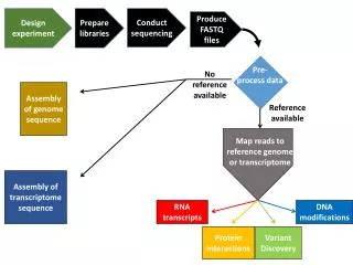
MR-scanner Raw data Scanner Subject Stimulus Delivery Reconstruction Distortion correction BOLD EPI Experiment design Co-registration Anatomy Function BOLD signal Statistical models Inference Func. & Anat. Group analysis
} Rich source of ideas for novel tools Scientific & Statistical Computing Core • Develop and implement new methodologies to meet user needs • Consult with IRP users/groups regarding • Experimental design • Processing methods and tools • Statistical inferences • Conduct classes on designing and processing FMRI experiments • Answer FMRI/MRI questions on message board • Distribute & maintain our open-source software tools • Facilitate cross-talk between different FMRI tools: • AFNI, FSL, FMRIstat, FreeSurfer, Caret, SPM, …
AFNI + SUMA • AFNI= collection of programs for FMRI analysis • Visualization • 2D, 3D, time-series, cortical surface (SUMA) • Time Series Analysis • Linear & nonlinear regression • Statistics on 3D Image Collections • 1-5 way ANOVA; non-parametrics; SEM • Data editing tools • Spatial and temporal filtering • 3D image registration • Clustering; ROI drawing & Atlas-based ROIs
The AFNI / SSCC Philosophy • Enable users to stay close to their data • Save intermediate results • Look at images and data in connected ways • User controls processing steps and parameters • Everyone has an opinion • Special problems need special solutions • Efficient (fast) implementations • Things that are easy and fast to do will get done more often • Help the users • Until our patience runs out
Next Set of Slides Features Added to AFNI and SUMA in Response to User Requests and/or Problems/Complaints (at least in part)
Feature: Atlases • Problem: Navigating in a complicated folded up 3D object (i.e., the brain) with few easily recognized landmarks • Solution: Coordinate-based brain atlases • Accepting the 5-10 mm uncertainty of brain coordinates • Atlas #1: Talairach-Tournoux atlas • As parsed by Peter Fox’s group at UT San Antonio • Atlas #2: Cytoarchitectonic atlases from Karl Zilles’ group at Forschungszentrum Jülich • 10 brains being sliced & diced & stained & scanned • About 40% complete at this time • Where Am I? + Jump To + Colorization + ROIs • Plans: keep up with Zilles; Animal atlases? …
A Feature: Skull Stripping • Problem: other skull stripping software (e.g., BET in FSL) didn’t work reliably enough • Solution was to re-visit problem from scratch, and build on BET’s surface growing algorithm • Then add new features: special knowledge about where the eyes are likely to be; 3D edges; etc. • Then test it on the hard cases from NIH (ab)users • Extra feature: extend it to monkey images • Plans: continue testing and improvements
A Feature: De-Spiking • Problem: occasional big spikes in echo planar images gathered for functional MRI • Problem eventually traced to gradient coil • In the meantime: can studies be saved? • Wrecks the standard time series analysis
Feature: Amplitude Modulated FMRI • Situation: Each stimulus event comes with an auxiliary parameter • May be measured (GSR, reaction time, …) or may be determined by experimenter • Want to determine if FMRI response magnitude is proportional to this auxiliary parameter • Solution was to add amplitude modulated regressors to AFNI’s 3dDeconvolve program • Two regressors per condition • First is: each stimulus response identical • Second is: each stimulus response proportional to auxiliary parameter for that stimulus • Plans: 2-3 params/event; event-wise amplitudes
Feature: Nonlinear Regression Models • Pharmacological models for time series analysis • AFNI’s nonlinear regression program 3dNLfim • Michaelis-Menton dynamics for BOLD FMRI with psychoactive drugs (e.g., ethanol) • Dynamic Contrast Enhanced MRI for quantifying Gd contrast leakage through blood-brain barrier
Feature: Smart Blurring • FMRI time series datasets are often smoothed (blurred) in space to • Reduce noise (by averaging) • Increase intra-subject activation “blob” overlap • Blurring brain & non-brain signals together is silly • When combining data from different scanners (i.e., multi-center studies), image smoothness varies • Should blur images until they have the same level of smoothness so that inter-scanner combinations make statistical sense • Developed a method for blurring inside a mask that stops when image noise reaches specified level of smoothness:
Feature: Structural Equation Modeling • SEM is a form of connectivity analysis • Input: correlations between activated ROIs • Regions where the activations fluctuate in strength together will be more highly correlated • Input: connectivity diagram between ROIs • Output: strength of connections • Can also search for “better” fitting connections
Feature: All-in-One Analysis Program • Common complaint: “AFNI is tooooooo hard to use” • Analysis of single subject data involves several steps, each instantiated in separate programs • Registration, smoothing, normalizing, model analysis • Solution is a program afni_proc.py that will run all these programs in a coherent sequence • Intermediate results are saved to make it possible to track backwards when results are confusing • This script is not intended to let the user avoid understanding the data analysis process!
Feature: Diffusion Tensor Analysis • Goal: Compute the Diffusion Tensor (etc.) from Diffusion Weighted image collections • Problem #1: log+linear method is inaccurate in highly anisotropic locations (the cool places to be) • Problem #2: published nonlinear solution methods not available in open-source software • Solution was to create and implement an efficient robust nonlinear method for finding the diffusion tensor D in each voxel • Also, a optional nonlinear image smoother (2D and 3D) to reduce noise in homogenous areas • Our code now incorporated into DTI Query, an open-source tractography program from Stanford
Feature: Inter-Modality Registration • Goal: Efficiently align 3D volumes acquired with different imaging contrasts • Solution is a general program using histogram-based measurements of image matching (e.g., mutual information) • This one is still very much a work-in-progress • Works pretty well on “simple” cases (e.g., whole-brain to whole-brain) • Dealing with partial-brain to whole-brain and with brain images that have holes in them is less reliable right now • Also want to add non-affine warping capabilities
A Example: Inter-Modality Registration Skull Stripped MRI … masked CT … CT overlaid on MRI in color - unaligned … CT overlaid on MRI in color - aligned
Feature: Analysis of Mn Contrast MRI • Mn is an MRI contrast agent and a calcium analog • Goal: time-dependent in vivo tract tracing in monkeys • Problems abound: • Like FMRI, signal changes are small • Other artifacts from day-to-day scanning are larger • Simple image subtraction isn’t reliable • Next 3 slides: some data and results …
A Mn Data: Different Days
A Mn Data: Subtract & t-Test
A Mn Data: Cleverer t-Test
Next Set of Slides Features Added to AFNI and SUMA in Response to Our Own Crazy Thoughts (mostly)
Realtime FMRI Functional activation& Motion estimation in realtime AFNI Dimon Feedback Receiver MR Scanner, Image Files
Movie capture from SUMA • Activation map projected from AFNI Surface-Based Analyses • Create cortical surface models, project 3D data to these surfaces, analyze in that space • Respects geometry and topology of cortex • Most AFNI statistical tools now work with image data defined over surfaces as well as over 3D volumes
NIfTINeuroimaging Informatics Technology Initiative • Goal: facilitate inter-operability of FMRI data analysis software • First fruit: NIfTI-1.1 standard for storing datasets defined over 3D volumes (plus time axis) • Works with AFNI, FSL, SPM, BrainVoyager, … • Agreement is not a one-time thing • Ongoing process is needed to deal with compatibility, extensions, new ideas along the same line, … • Efforts underway: • NIfTI-G: standard for storing cortical surface models (and associated data) • NIfTI-W: standard for storing non-affine spatial warps
Closely Linked Communication • Programs “talk” to each other (esp. AFNI & SUMA) • Exchange data • Issue commands - you can script many parts of the AFNI & SUMA graphical interfaces AFNI SUMA 3dSkullStrip
Developer-friendliness Realtime physiological monitoring using AFNI: Jerzy Bodurka, FIM/LBC/NIMH
L R Brain State Classification • Train Support Vector Machine (SVM) classifier on a collection of pre-categorized 3D brain images • e.g., “looking at house” and “looking at face” • Classifies new 3D images into the categories From Stephen LaConte; Emory, transitioning to Rice
Penultimate Slide • Much of our most fruitful and satisfying work comes from close and ongoing interactions with investigators that have interesting problems • Derived from studies that are pushing the envelope of deriving information from MRI • We are here to provide solutions to problems (of image analysis) • Your current short-term problems (lots of these!) • Your actual longer-term problems • What we think your future needs will be
Ultimate Slide The Team