ANGIOGENESIS IMAGING METHODS
ANGIOGENESIS IMAGING METHODS. 성균관의대 핵의학교실 이 경한. AREAS OF DISCUSSION. What is Angiogenesis ?. Why Angiogenesis Imaging ?. Current Methods are Available ?. Prospects of Future Techniques. WHAT IS ANGIOGENESIS ?. ANGIOGENESIS. “The growth of new blood vessels”


ANGIOGENESIS IMAGING METHODS
E N D
Presentation Transcript
ANGIOGENESIS IMAGING METHODS 성균관의대 핵의학교실 이 경한
AREAS OF DISCUSSION • What is Angiogenesis ? • Why Angiogenesis Imaging ? • Current Methods are Available ? • Prospects of Future Techniques
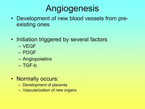
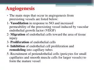
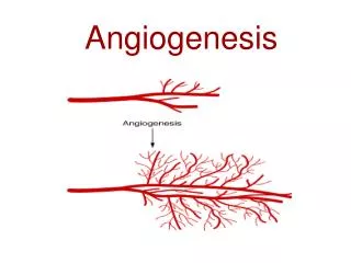
ANGIOGENESIS • “The growth of new blood vessels” • An important natural process occurring in the body, both in health and in disease. • Occurs in healing wounds and for restoring blood flow to tissues after injury. • The healthy body controls angiogenesis through a series of "on" and "off" switches: - “on” switch: angiogenesis growth factors - “off” switch: angiogenesis inhibitors
Excessive Angiogenesis • Occurs in cancer, DM blindness, macular degen, RA, psoriasis, … • Occurs when ds. cells produce abnormal amounts of angiogenic GFs, overwhelming the effects of natural angiogenesis inhibitors • Feed ds. tissues, destroy normal tissues, and allow tm metastasis Insufficient Angiogenesis • Occurs in coronary artery disease, stroke, and delayed wound healing • When the tissue cannot produce adequate amounts of angiogenic GFs • Lead to to improper circulation and tissue death
Angiogenesis Facts • > 19 known angiogenic growth factors • > 5 angiogenic GF being tested in humans to heal wounds • > 30 known natural angiogenesis inhibitors found in the body. • > 300 angiogenesis inhibitors have been discovered to date. • > 184 million could benefit from antiangiogenic therapy • > 314 million would benefit from pro-angiogenesis therapy • > 6,500 cancer pts have been treated with antiangiogenic therapy • > 1,000 heart ds. pts received experimental angiogenic therapy • > $4 billion invested in R&D angiogenesis-based medicines
Angiogenic Cascade • Tm interaction with vasculature switch to angiogenic phenotype, enabling tumor progression - Endothelial receptor binding / activation - Formation of angiogenic mother vessels - Morphogenesis of mother vessels - Basement membrane dissolution - Endothelial cell proliferation - Endothelial cell migration - Vascular tube formation - Arterial-venous differentiation - Vascular stabilization
Known Angiogenic Growth Factors • Angiogenin • Angiopoietin-1 • Del-1 • Fibroblast growth factors • Follistatin • Granulocyte colony-stimulating factor (G-CSF) • Hepatocyte growth factor /scatter factor • Interleukin-8 (IL-8) • Leptin • Midkine • Placental growth factor • Platelet-derived endothelial cell GF • Platelet-derived growth factor-BB (PDGF-BB) • Pleiotrophin (PTN) • Proliferin • Transforming growth factor-alpha (TGF-alpha) • Transforming growth factor-beta (TGF-beta) • Tumor necrosis factor-alpha (TNF-alpha) • VEGF / vascular permeability factor
Lessons from Early Clinical Trials • Animal studies do not directly translate to human studies • Host responder characteristics remain poorly understood • Combination therapy may enhance clinical outcome • Conventional oncology trial strategies require modification • Require new standards rather than change in tumor mass for monitoring thx response
Workshop on Angiogenesis Imaging Methodology Sponsored by the Biomedical Imaging Program of the NCI, 2000 • Experts from imaging modalities: US, CT, MRI, PET/Scan • Controversy over the adequacy of present anatomical imaging measurements and definition of response criteria • Challenges from growing number of antiangiogenesis clinical trials • Interest in imaging techniques that can provide an early indicator of effectiveness at a functional or molecular level has increased
Prognostic Value of Tumor Angiogenesis Assessment • Gastric carcinoma. Erenoglu C, Dig Surg. 2000 • Esophageal cancer. Millikan KW, Am Surg. 2000 • Node-positive Breast cancer. Viens P, Breast Cancer Res Treat. 1999 • Tumour angiogenesis and prognosis. Morgan KG, Histopathology. 1998 • Tumor angiogenesis in prognosis. Fox SB, Invest New Drugs. 1997 • Breast cancer. Goulding H, Hum Pathol. 1995 • Bladder cancer. Bochner BH, J Natl Cancer Inst. 1995 • Squamous cell carcinoma. Zatterstrom UK, Head Neck. 1995 • Bladder carcinomas. Dickinson AJ, Br J Urol. 1994
Present Gold Standard Immunohistochemistry • Invasive • Repeated exam difficult • Large variation • Sampling error Anti-CD31
CT Methods • Performed with contrast agents to define the intravascular compartment, including blood flow, blood volume, transit time, and capillary permeability. • Functional CT techniques can delineate increases in tissue perfusion Ultrasound Methods • Can identify vascular features at resolution of 50-200 um vessels • Contrast-enhancement using an intravascular agent can generate an index of blood flow, blood volume, or vascularity within tumor • Targeted imaging using ultrasound destruction of microbubbles may provide even further resolution of the tumor vascular tree.
MRI Methods • Can define blood volume and permeability using dynamic enhancement of blood pool contrast agents. • Gadolinium-DTPA can distinguish between normal versus malignant leaky tissues, reflecting the hyperpermeable tumor vasculature. • Contrast uptake also correlates with microvessel density in experimental tumors.
Gene Therapy with VEGF for Inoperable CAD JM Isner, The Annals of Thoracic Surgery, 1999 SPECT METHODS
Gene Therapy for Myocardial Angiogenesis. Initial Clinical Results with Direct Myocardial Injection of phVEGF165 as Sole Therapy Pre Post Stress Rest JM Isner, Circulation1998
Catheter-Based Myocardial Gene Transfer for Angiogenesis JM Isner, Circulation2001
PET METHODS • Evaluates tumor metabolism, as well as blood flow and volume. • H2015, 11CO, and 18FDG, characterizes neoplastic tissue • Antiangiogenic agents should diminish blood flow and decrease tumor metabolism • Radiolabeled fluoromisonidazole (FMISO) has been used to quantitate hypoxia in the rat glioma by PET and may provide functional information about the results of antiangiogenic therapy.
Future Prospects For Nuclear Imaging Methods
Requirements of Angiogenesis Imaging Methods • Measure vascularity and its change with high SN and SP • The small size of microvessels precludes direct visualization by conventional angiography • Cases often have late-stage disease and a heavy tumor burden with an extensive established vascular supply: imaging must accurately quantify small changes against a potentially large signal background • As antiangiogenic therapy may require lifelong treatment, a non-invasive and costeffective technique would be highly desirable.
Guidelines for Nuclear Imaging Developement • Effect of anti-angiogenic drugs on parameters measured by nuclear imaging has not been evaluated • Techniques currently being used in ongoing clinical trials of anti-angiogenic drugs be studied in animal models to evaluate the changes induced by anti-angiogenic therapy. • New approaches include integrins, annexin V, hypoxia agents, proliferative indices, and various receptor ligands
Integrin v3 [18F]Galacto-RGD Integrin Imaging Strategies • The v3 integrin is expressed on newly formed endothelial cells • and is thought to anchor the new blood vessel in the tumoral stroma • Extracellular matrix proteins interact to v3 integrin via RGD sequence
Table 1 Biodistribution data for [18F]Galacto-RGD in melanoma-bearing nude mice and osteosarcoma-bearing mice v v Time (min) Osteosarcoma (ß3 positive) Melanoma (ß3 positive) Melanoma (negative) Blood 10 2.58 ± 0.41 2.93 ± 0.53 4.70a 120 0.13 ± 0.03 0.05 ± 0.01 0.13 ± 0.07 Liver 10 5.47 ± 0.11 5.64 ± 2.27 6.40 120 1.97 ± 0.27 1.25 ± 0.05 1.79 ± 0.58 Kidneys 10 8.67 ± 0.52 8.48 ± 0.84 10.82 120 2.21 ± 0.46 1.52 ± 0.10 1.86 ± 0.46 Muscle 10 0.80 ± 0.13 1.13 ± 0.18 1.44 120 0.28 ± 0.02 0.15 ± 0.02 0.18 ± 0.03 Tumor 10 2.88 ± 0.32 3.90 ± 1.36 1.88 120 1.68 ± 0.49 1.49 ± 0.10 0.44 ± 0.24 Lung 10 3.43 ± 0.24 3.64 ± 0.52 4.84 120 0.72 ± 0.15 0.53 ± 0.05 0.71 ± 0.24 Colon 10 3.77 ± 0.16 1.74 ± 0.55 1.68 120 1.59 ± 0.46 3.53 ± 0.44 0.76 ± 0.27 Biodistribution in Melanoma M21() and M21-L() Bearing Mice Biodistribution in Tumor Bearing Mice
Haubner R, Cancer Res 2001 [18F]Galacto-RGDPET of Melanoma Bearing Mice v Dose-dependent blockade of uptake by selective pentapeptide
SYNTHESIS OF 18F-FLUOROPROPYLSQUALAMINE AS ANGIOGENESIS IMAGING AGENT • Squalamine inhibits angiogenesis and solid tumor growth in vivo • N-fluoropropylsqualamine has similar activities as squalamine • F-18 N-fluoropropylsqualamine was synthesized in 4-7% yield SNM meeting, 2001 C-Y Shiue*, Univ. Penn and Magainin Pharmaceuticals Inc.
Guidelines for Nuclear Imaging Developement • Effect of anti-angiogenic drugs on parameters measured by nuclear imaging has not been evaluated • Techniques currently being used in ongoing clinical trials of anti-angiogenic drugs be studied in animal models to evaluate the changes induced by anti-angiogenic therapy. • New approaches include integrins, annexin V, hypoxia agents, proliferative indices, and various receptor ligands • Anti-angiogenic drugs themselves be radiolabeled to directly study the pharmacokinetics of the drug.
Therapeutic Targets in Tumor Angiogenesis Growth factor antagonists - Inhibition of angiogenic factor production - Anti-growth factor ribozymes - Soluble growth factor receptors - MoAb against angiogenic factors Endothelial signal transduction inhibition - Receptor tyrosine kinase inhibition - Protein kinase C inhibition Inhibitors of endothelial cell proliferation - Cell-cycle inhibitors Matrix metalloproteinases inhibition - Selective inhibitors of MMP-2, MMP-9 - Non-selective MMP inhibition Endothelial surface marker targeting - Anti-integrin antibodies or cyclic peptides Endothelial cell subpopulation inhibitors - Suppression of endothelial progenitor cells Endothelial cell destruction - Vascular targeting agents
Known Angiogenesis Inhibitors • Antiangiogenic antithrombin III • Cartilage-derived inhibitor (CDI) • CD59 complement fragment • Platelet factor-4 (PF4) • Prolactin 16kD fragment • Proliferin-related protein (PRP) • Fibronectin fragment • Gro-beta • Heparinases • Heparin hexasaccharide fragment • hCG • Interferon alpha/beta/gamma • Interferon inducible protein (IP-10) • Interleukin-12 • Angiostatin (plasminogen fragment) • Kringle 5 (plasminogen fragment) • Endostatin (collagen XVIII fragment) • Metalloproteinase inhibitors • 2-Methoxyestradiol • Placental ribonuclease inhibitor • Plasminogen activator inhibitor • Retinoids • Tetrahydrocortisol-S • Thrombospondin-1 (TSP-1) • TGF-b • Vasculostatin • Vasostatin (calreticulin fragment)
123I-Taxol for Angiogenesis Imaging 7000 3 uCi 6000 6 uCi 5000 4000 CPM/ug/ml protein 3000 2000 1000 0 0 50 100 150 Time (min) YS Choe, 1999
Targeted Angiogenesis Tumor Vascular Imaging With Radiolabeled Endostatin • 131I-labeled endostatin and 99mTc-labeled endostatin • In tumor-bearing rats, Tm/tissue count ratios incr. with time • Tumor %ID/g was 0.2-0.5 for 99mTc and 0.2-1.2 for 131I • Images visualized tumor clearly with radiolabeled endostatin • 99mTc-EC-endostatin could assess treatment response SNM meeting, 2001 D. J. Yang,. MD Anderson and EntreMed
ANGIOSTATIN • Angiostatin is a proteolytic fragment of plasminogen • A potent inhibitor of angiogenesis and tumor growth
300 250 200 Eluted Activity (Ci) 150 100 50 0 0 5 20 10 15 Time (tube number) 50 kD 123I-Angiostatin Synthesis KH Lee, 2001
(%) 100 80 Activity in serum Activity in clot 60 80 40 70 % total activity 60 20 50 0 40 0 40 80 120 160 200 30 minutes 20 10 0 5min 10 min 20 min Blood Clearance Intravascular Activity of 123I-Angiostatin KH Lee, 2001
9.0 8.0 7.0 6.0 5.0 4.0 3.0 2.0 1.0 0.0 Blood Heart Lung Liver Spleen Pancr. Tumor muscle Kidney Tumor Uptake in Colon Cancer Bearing Mice KH Lee, 2001
Tumor/Contralateral 6.0 Tumor/Lung Tumor/Liver 5.0 Tumor/Heart 4.0 Count Ratio 3.0 2.0 1.0 0.0 20 30 40 50 60 70 80 90 100 110 120 Minutes Sequential Tumor Uptake Ratio in Image ROIs KH Lee, 2001
SUMMARY • Rapidly accumulating knowledge of tumor angiogenesis is providing critical insights into new opportunities for imaging • Angiogenesis imaging is critical for optimizing antiang. therapy • Conventional techniques may be adapted to measure blood flow, blood volume, permeability, microvessel density, and tissue metabolism • Future approaches for imaging angiogenesis per se will likely exploit the molecular features of new blood vessel growth. • Novel imaging targets include cell surface integrins, endothelial apoptosis, angiopoietins and other signatures of angiogenesis. • These new modalities will help create a platform for bringing antiangiogenic cancer therapy into standard oncology practice
FUTURE DIRECTIONS • Many questions remain about the angiogenic process and how it is regulated. And antiangiogenic imaging methods now in development face uncertainties of efficiency • Despite the obstacles, angiogenesis imaging offer the promise of an additional diagnostic modality for our current armamentarium. • Angiogenesis imaging may turn out to have significant benefits because they target easily accessable cells and are unaffected by resistance. • These imaging methods may also be used to evaluate other diseases characterized by abnormal angiogenesis, such as ischemic disease, arthritis, and benign tumors. • Clearly, then, antiangiogenic drugs have exciting potential as therapies for a number of serious conditions-in addition to cancer.
TUMOR TARGETING WITH RADIOLABELED INTEGRIN aVb 3 BINDING RGD PEPTIDES IN A NUDE MOUSE TUMOR MODEL M. Janssen, Dupont Pharmaceuticals • 111In and 99mTc-labeled RGD peptide in ovarian cancer bearing BALB/c mice • Maximal tumor uptake of 7.5 %ID/g of 111In-labeled peptide at 2 hrs pi. • Tumors were clearly visualized by gamma camera scintigraphy. • Tumor growth was significantly delayed after injection of 90Y-RGD peptide • RGD-peptide labeled with either 111In or 99mTc specifically localizes in human tumor xenografts and in various normal tissues in nude mice. Labeled with 90Y this peptide has potential for peptide receptor radionuclide therapy.
True Angiogenesis Inhibitors a) Specific inhibitors of angiogenic growth factors - Angiozyme (Ribozyme Pharmaceuticals) - Avicine (AVI Biopharma) - Suramin (NCI) - rhu MabVEGF (Genentech) b) Inhibitors of growth factor-receptor binding - IMC-1C11 (ImClone) - IM862 (Cytran) - PI-88 (Progen Industries) c) Specific tyrosine kinase inhibitors - PTK787 (Novartis) - SU5416 (SUGEN) - SU6668 (SUGEN) d) Anti-endothelial proliferative agents - TNP-470 (TAP Pharmaceuticals) e) Anti-integrin agents - EMD121974 (Merck KgaA) - Vitaxin MedImmune f) Inhibitors of angiogenic factor production - Octreotide (Novartis) g) Upregulators of angiogenesis inhibitors - ImmTher (Endorex) h) Unknown mechanism - Angiostatin (EntreMed) - Endostatin (EntreMed)
Nonselective Antiangiogenic Agents a) Low-dose cytotoxic chemotherapy drugs - Cyclophosphamide, 5-Fluorouracil - Methotrexate, Vinblastine b) Matrix metalloproteinase inhibitors - BMS275291 (Bristol-Myers Squibb) - Captopril (Bristol-Myers Squibb) - Col-3 (CollaGenex) - Marimastat (British Biotech) - Neovastat (Aeterna Laboratories) - Prinomastat (Agouron Pharmaceuticals) - Solimastat (British Biotech) c) Anti-cytokine agents - Thalidomide (Celgene Corp.) - CC 4047 (Celgene Corp.) - CC 5013 (Celgene Corp.) - CC 7085 (Celgene Corp.) - CDC801 (Celgene Corp.) d) Cox-2 inhibitors - Celecoxib (GD Searle) e) Anti-tubulin agents - Paclitaxel (Angiotech) f) Cell locomotion inhibitors - Interferon alfa2a (Hoffman-LaRoche) g) Ion flux inhibitors - Carboxyamidotriazole (NCI) h) Anti-mitochondrial agents - Apra (Cell Therapeutics) i) Nonspecific tyrosine kinase inhibitors - Flavopiridol (NCI) - Genistein (Amino A) j) Copper-lowering agents - D-Penicillamine (NCI) - Tetrathiomolybdate (University of Michigan) k) Cell cycle inhibitors - Ro 317453 (Roche) Vascular Targeting Agents a) Anti-tubulin agents - Combretastatin A4 Prodrug (OXiGENE) b) Ion transport inhibitors - Squalamine (againin Pharmaceuticals) c) Receptor-driven inducers of endothelial apoptosis - CM101 (arboMed)
Desirable Characteristics of Radiotracers for Angiogenesis Imaging • Widely available • Fully validated • Highly sensitive to changes in the biochemical process • Biochemical parameters can be extracted from a single scan
Classification of Angiogenesis Inhibitors • True angiogenesis inhibitors • Halt only vascular sprouting and do not destroy preestablished tumor blood vessels. • Generally slow tumor growth within several days to a week or more. • Expected effect is disease stabilization rather than tumor regression • Vascular targeting agents • Destroy the pre-existing tumor vasculature. • In animal studies, effect is observable within hours. • Acute endothelial cell death, thrombosis, and tumor mass hypoxia and necrosis result. • Non-selective antiangiogenic agents • Antiproliferative or cytotoxic effects on multiple cell types as well as endothelium. • Dose adjustment, schedule, or delivery mode may produce anti-endothelial effects.
The Angiogenesis Process: How Do New Blood Vessels Grow? • Diseased tissue releases angiogenic GFs that diffuse into the nearby tissues • The angiogenic GFs bind to specific Rp on the EC of nearby preexisting blood vessels • Once GFs bind to their Rps, the ECs become activated. Signals are sent from the cell's surface to the nucleus. The endothelial cell's machinery begins to produce new molecules including enzymes • Enzymes dissolve tiny holes in the basement membrane surrounding all existing blood vessels • The endothelial cells begin to divide and migrate out through the dissolved holes of the existing vessel towards the diseased tissue or tumor • Specialized molecules called adhesion molecules, or integrins serve as grappling hooks to help pull the sprouting new blood vessel sprout forward • Matrix metalloproteinases are produced to dissolve the tissue in front of the sprouting vessel tip in order to accommodate it. As the vessel extends, the tissue is remolded around the vessel • Sprouting endothelial cells roll up to form a blood vessel tube • Individual blood vessel tubes connect to form blood vessel loops that can circulate blood • Finally, newly formed blood vessel tubes are stabilized by specialized muscle cells (smooth muscle cells, pericytes) that provide structural support. Blood flow then begins