Download

1 / 50
510 likes | 1.27k Vues
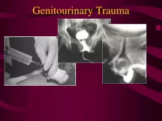
Genitourinary Trauma in the Emergency Department. Tran Duc Lai, MD Hue University Hospital James Ramseier , MD Las Vegas, USA. Epidemiology. Injury frequency of abdominal organs: S plenic injuries—25% Liver injuries—15-20% Renal Injuries—10% Most common between 20-35 years of age

E N D
Genitourinary Trauma in the Emergency Department Tran Duc Lai, MD Hue University Hospital James Ramseier, MD Las Vegas, USA
Epidemiology • Injury frequency of abdominal organs: • Splenic injuries—25% • Liver injuries—15-20% • Renal Injuries—10% • Most common between 20-35 years of age • More common in men • Mechanism • Blunt—90% • Penetrating—10%
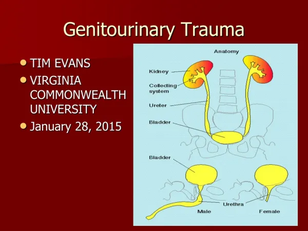
Anatomy • Upper Urinary Tract • Kidney • Ureter • Lower Urinary Tract • Bladder • Urethra
Prehospital Care • No specific intervention is needed • One exception… • Penile amputation • Direct pressure to stop bleeding • Wrap amputated penis in sterile dressing • Place in container of ice– not in direct contact with ice
History • GU tract assessed during secondary survey • Mechanism of Injury—blunt vs. penetrating • Past medical history • Problems with renal function • High risk for renal insufficiency (diabetes, hypertension • Uninephric • Gross hematuria • Difficulty voiding since injury
Physical Exam • Flank exam • Risk stratifies injury to kidneys • Tenderness, palpable mass, ecchymosis • Costal margin exam • Injury to kidneys, spleen, and liver • Thoracolumbar spine exam • Palpation to identify potential fractures • Transverse spine fracture higher risk of renal injury • Back exam for ecchymosis • Pelvic stabilility • Instability increases risk of bladder injury
Physical Exam • Rectal exam • May not be useful in all trauma patients • Reserved for patients with: • Pelvic fractures • Evaluates prostate: high riding/boggy? urethral injury • Penetrating wound to lower abdomen • Evaluate rectal tone when spinal cord injury suspected • Perineal exam • Hematoma and edema • Suggests possible urethral injury • External genitalia exam • Penile injury: edema, laceration, deformity • Urethral injury: blood at meatus • Testicular injury: edema, laceration, deformity
Foley Catheter Placement • Indications in critical patient: • Test for hematuria • Monitor urine output • Ease of urination • Contraindications Retrograde urethrogram • Blood at urethral meatus • Boggy/high-riding prostate • Perinealhematoma • Difficulty catheterization Retrograde urethrogram • Urethral injury • Urethral stricture • Prostatic hypertrophy • Cancer
Urinalysis • GU trauma screening test UA for hematuria • Urine b-hCG for pregnancy status • Gross hematuria • Renal injuries 50% • Renal injuries without hematuria 5% • Renal artery injury • Disruption of the uteropelvic junction • Evaluate GU tract and other abdominal organs • CT scan with delayed images: kidneys +abd organs • Cystogram: bladder
Urinalysis • Microscopic hematuria • Pediatric population • Routinely used but controversial • Cutoff levels of 5-50 RBC/hpf used for additional abdominal imaging • Vast majority of GU injuries have >50 RBC/hpf • Adult population • Controversial • Cutoff levels of >10 –>25 RBC/hpf used for additional abdominal imaging
Urinalysis • Microscopic hematuria • Adult population • Standard based on old studies that attempted to identify renal injury requiring intervention • Blunt trauma patient needs upper urinary tract imaging if… • Gross hematuria • Microscopic hematuria and shock • These studies did not take in to account: • Renal injuries not requiring intervention • Ability of microscopic hematuria to account for non-GU injuries • The ability of microscopic hematuria to detect lower urinary tract injuries • Degree of hematuria requiring abdominal imaging varies from >10 RBC/hpf to >25 RBC/hpf • Consider the whole picture: degree of hematuria, mechanism of action, and additional physical exam findings
Additional Labs • Complete blood count • Type and screen • BUN and Creatnine levels • Increase with: • Renal injury • Ureter/bladder injury • CT abdomen with contrast • X-ray • Ultrasound
Specific Injuries & Treatment • Upper urinary tract • Renal injuries • Lumbar spine fractures (esp. transverse process fractures) • Injury grade guides treatment • Grade I-III: non-operative repair in children and adults • Grade IV: half require surgery • Grade V: all require surgery • Acute complications • Hemorrhage, urinoma, hydronephrosis, arterial pseudoaneurysm, infection • Long-term complications • Renal failure, hypertension
Specific Injuries & Treatment • Renal injuries (cc) • Renovascular injuries • Most severe, but rare • Renal artery thrombosis most common • CT abdomen with contrast = screening test of choice • Renal pedicle contrast extravasation • Complete/focal non-enhancing kidney • Renal hilarhematoma • Hilar hematoma + normally enhancing kidney => renal vein injury
Specific Injuries & Treatment • Renovascular injuries (cc) • Treatment • Complete devascularization • Emergency— vascular repair attempted; most result in nephrectomy • Renal artery pseudoaneurysm/dissection • Angiographic embolization • Endovascular stent placement
Renal Injury Grade 4
Renal Injury Grade 1; contusion
Renal Injury Cortical laceration >1 cm Grade 3
Renal Injury Grade 1 Subcapsular hematoma
Renal Injury subcapsular and perinephric hematoma Grade 2
Renal Injury Renal laceration >1 cm Grade 3
Renal Injury Shattered kidney with large hematoma Grade 5
Renal Injury Grade 4 Segmental infarction
Renal Injury Grade 5 Devascularization
Renal Injury Grade 3 renal laceration Normal one-shot IVP
Renal Injury Grade 3 renal laceration Absent right nephrogram
Renal Injury Grade 5 injury; shattered right kidney Absent right nephrogram
Specific Injuries & Treatment • Upper urinary tract (cc) • Ureteral injuries • Rare, 1% of GU injuries • Gunshot wounds 94% • Stab wounds 5% • Blunt trauma 1% • Sudden decelerating force • No specific physical exam findings • Hematuria 75% • Screening: CT abdomen with contrast and delay views • Grading: scale not well accepted • Diagnostic: Retrograde pyelography • Treatment: ureter with active extravasation of urine • Stenting, primary closure, ureteroneocystostomy • Ureteroureterostomy less frequent • Complications: urinoma, abscess, stricture, hydronephroma, fistula, ileus
Specific Injuries & Treatment • Lower urinary tract • Bladder injuries • Classic triad: gross hematuria, suprapubic pain, inability to void • Risk of injury increased • Pelvic fractures, pregnant (>1st trimester,) intoxicated • Hematuria • Gross hematuria in 67% • Injury rare when <25 RBC/hpf • Diagnosis • Bladder CT scan • Plain film cystogram • Delays may show increase in BUN & Creatnine
Specific Injuries & Treatment • Bladder injuries • Injury classification and treatment • Contusion/hematoma • Treatment: observe for resolution of hematuria • Intraperitoneal rupture • Sudden increase in intra-bladder pressure • Treatment: operative repair + urine catheter for 14 days + broad spectrum antibiotics • Repeat cystogram looking for urine leak • Catheter removed if no leak • Extraperitoneal rupture • Bony fragment laceration; sheering force • Treatment: observe for 7 days + urine catheter • Evaluation for resolution of hematuria • No hematuria repeat cystogram • Complications: infection, urinoma, inability/difficulty voiding, fistula • Emergency department visit post-op with urinary retention • Place urine catheter after consulting urologist • Check urine for infection
Bladder Injury Extraperitoneal rupture
Bladder Injury Intraperitoneal rupture
Bladder Injury Extraperitoneal rupture
Bladder Injury Intraperitoneal rupture
Bladder Injury Extraperitoneal rupture
Bladder Injury Intraperitoneal rupture
Specific Injuries & Treatment • Urethral injuries • Rare; male 95% • Risk increased with • Pelvic fractures: pubic symphysis and sacroiliac diastasis, straddle fracture, Malgaigne fracture • Straddle injuries • Penetrating trauma close to urethra • Penile fracture • Physical exam: • Blood at urethral meatus, perinealhematoma/edema, high riding prostate on rectal—rare • Gross hematuria—common
Specific Injuries & Treatment • Urethral injuries (cc) • Gross hematuria + difficulty placing catheter => urethral evaluation • Extension of urethral laceration, increase bleeding, contamination of hematoma • Grading system exists but is not well accepted • Injury severity determined by: • Location of the injury • Complete vs incomplete transection of the urethra • Diagnosis: Retrograde urethrogram • Treatment: if urethra is injured, consult urology
Urethral Injury Normal
Urethral Injury Partial urethral disruption Complete urethral disruption
Specific Injuries & Treatment • External genitalia injuries • Penile laceration • Superficial penile laceration repair in emergency dept. • Complex penile laceration consult urology • Compromised Buck’s fascia consult urology • Penile fracture • Occur during coitus or masturbation • Presentation: penile pain, loss of erection, deformity, swelling, difficulty voiding • Treatment: • Involvement of corpus caverosum or Bucks fascia (common) operative intervention • Superficial to Bucks fascia (less common) nonoperative management
Specific Injuries & Treatment • External genitalia injuries (cc) • Penile amputation • Treatment: reimplantation if warm ischemic time <6 hours • In emergency department, penis is kept cold (wrapped in dry gauze on ice,) and urology consulted immediately • Penile foreign bodies • Blunt foreign bodies in the distal urethra removal in the emergency department • Sharp, not easily visualized in the proximal urethraurology consultation for operative intervention • Complication: strictures, impotence, incontinence
Specific Injuries & Treatment • Testicular injuries • Uncommon; blunt trauma 85% • Testicular rupture: most serious injury • Hematocele: hemorrhage in to the tunica vaginalis • Testicular dislocation: rare; manual reduction in the emergency department recommended • Diagnosis: Ultrasound study of choice • Treatment • Nonoperative intervention for minor injuries • Scrotal elevation, ice, non-steroidal antiinflammatory drugs, limited activity • Operative intervention • Testicular rupture • Expanding hematocele • Inability to reduce testicular dislocation • Scrotal degloving • Testicular salvage as low as 40% • Complications: bleeding, abscess, skin necrosis, infertility, testicular atrophy, testicular necrosis
Testicular Injury Injured or uninjured?
Summary • Kidneys are the 3rd mostcommonlyinjured abdominal organ • Specific GU trackinjuryisevaluatedduring the secondarysurvey • Most renal injuries are minorwith no long term complications • Gross hematuria OR microhematuria + shock = GU trauma • Renovascular injuries require prompt diagnosis and urological consultation • Pelvic fracture + >25 RBC/hpf => considerbladderinjury • Blood aturethralmeatus, perinealhematoma, high-riding prostate => considerurethralinjury • Imaging selection: • Renal CT abdomen withdelayed images; one-shot IVP; ultrasonography • Ureteral one-shot IVP • Bladder plain film cystogram; CT cystogram • Urethral retrogradeurethrogram • Testicular ultrasound