Metalloproteins in Oxygen Reactions: Functions and Significance
330 likes | 482 Vues
Explore the crucial role of metalloproteins in oxygen reactions in aerobic organisms, including energy production, detoxification, and oxygen transport mechanisms. Learn about the kinetic and solubility challenges faced by organisms in utilizing oxygen and how metalloproteins address these issues with transition metal complexes. Discover the activation of oxygen for reactions with organic molecules through metal-oxygen adduct formation. Dive into the functions of oxygen transport proteins and oxygenases in facilitating biological processes. Gain insights into the structure and function of metalloproteins in oxygen reactions.

Metalloproteins in Oxygen Reactions: Functions and Significance
E N D
Presentation Transcript
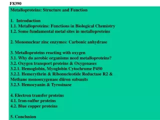
F8390 • Metalloproteins: Structure and Function • Introduction • 1.1. Metalloproteins: Functions in Biological Chemistry • 1.2. Some fundamental metal sites in metalloproteins • 2. Mononuclear zinc enzymes: Carbonic anhydrase • 3. Metalloproteins reacting with oxygen • 3.1. Why do aerobic organisms need metalloproteins? • 3.2. Oxygen transport proteins & Oxygenases • 3.2.1. Hemoglobin, Myoglobin Cytochrome P450 • 3.2.2. Hemerythrin & Ribonucleotide Reductase R2 & • Methane monooxygenase diiron subunits • 3.2.3. Hemocyanin & Tyrosinase • 4. Electron transfer proteins • 4.1. Iron-sulfur proteins • 4.2. Blue copper proteins • 5. Conclusion
3. Metalloproteins reacting with oxygen 3.1. Why do aerobic organisms need metalloproteins? Cytochrome P450 Cells of aerobic organisms need oxygen. First, oxygen is needed to gain energy from food (respiration) and for other processes. Second,toxic organic substances are eliminated from the body by oxidation, whereupon OH-groups are attached to the molecule (this specific process is called hydroxylation, in mammals it occurs mainly in the liver). This renders the toxic molecule water-soluble and it can be eliminated (through the urine in mammals). Cellular respiration C6H12O6 + 6 O2 6 CO2 + 6 H2O DG0 = -674 kcal/mol Elimination of xenobiotics. Example: hydroxalation of hexane by Cytochrome P450 minor minor major
Use of oxygen by aerobic organisms is hampered by two problems: 1. The solubility problem Water solubility of oxygen at 25oC and pressure = 1 bar is at 40 mg/L water. This is not enough to guarantee the oxygen supply to mitochondria by mere diffusion. Cells of aerobic organisms use therefore oxygen transporters. 2. The kinetic problem Oxygen has two unpaired electrons in its ground state and forms therefore a triplet state. The overwhelming majority of organic molecules (such as glucose or n-hexane) have all electrons paired and occur therefore in the singlet state. The products of oxidation of organic molecules, CO2 and H2O, are also in singlet states. According to the so-called Wigner-rule, processes in which the spin-state changes are « spin-forbidden », that is, they have a large kinetic barrier. The solution of the problem is binding of O2 to a transition metal complex. In transition metal complexes, spin-state changes are less inhibited due to the spin-orbit coupling. The oxygen-bound metal complex can therefore transit from a triplet state to a singlet state, and then react with an organic substrate which has also a singlet ground-state.
Activation of O2 with the help of a transition metal complex: Adduct formation from a pentacoordinated [FeL5]2+ complex and O2
2p 2p 2s 2s O O2 O Molecular orbital level diagram for O2: 3Sg- state
y x z Bonding and antibonding MOsformed by AOs 2p in the O2 molecule s*2p p*2ph antibonding xz p*2pv yz p2ph xz bonding p2pv yz s2p
y x z O O Bonding and antibonding MOsforming a p bondin the O2 molecule p*2ph p*2pv antibonding bonding p2ph p2pv O O
Activation of O2 with the help of a transition metal complex: Adduct formation from a pentacoordinated [FeL5]2+ complex and O2
z2 x2-y2 xy xz yz Splitting of d orbitals in an octahedral environment (6 equal ligands) Cetral transition metal atom Lone-pairs of ligands M
z2 x2-y2 xy xz yz Splitting of d orbitals in an tetragonal environment (5 equal ligands) Cetral transition metal atom Lone-pairs of ligands 6 ligands octahedral field 5 ligands octahedral field xz M
z2 x2-y2 xy xz yz Splitting of d orbitals in an tetragonal environment (5 equal ligands) Cetral transition metal atom Lone-pairs of ligands 6 ligands octahedral field 5 ligands octahedral field x2-y2 z2 xz M
Remember for next slide z2 x2-y2 xy xz yz xz Splitting of d orbitals in an tetragonal environment (5 equal ligands) FeII is a d6 ion Cetral transition metal atom Lone-pairs of ligands 6 ligands octahedral field 5 ligands octahedral field x2-y2 z2 xz xy xz M
From last slide dz2 empty 3O2 + 1[L5Fe] 3[L5FeO2] spin-allowed: n° of unpaired electrons unchanged 3O2 One of the p* orbitals of O2 overlaps with the dz2 orbital of Fe and forms a bond; the other p* orbital is non-bonding 1[L5Fe] (only the two unpaired valence electrons shown)
3O2 + 1[L5Fe] 3[L5FeO2] spin-allowed: n° of unpaired electrons unchanged 3O2 One of the p* orbitals of O2 overlaps with the dz2 orbital of Fe and forms a bond; the other p* orbital is non-bonding Stabilization! 3[L5FeO2] 1[L5FeO2] 1[L5Fe] process spin-forbidden but rendered possible by spin-orbit coupling (only the two unpaired valence electrons shown)
In transition metal complexes, spin-orbit coupling renders spin-forbidden transitions possible. Metal complexes can therefore activate (triplet) oxygen for reactions with (singlet) organic molecules. + 1[Substrate] 1[MLn O2]m+ [MLn]m+ + 3O2 1[Oxidation products] Metal-oxygen adducts can also be used as oxygen carriers! 2. Oxygen transport proteins & oxygenases
Oxygen transport proteins: O2 binding in active sites Hemoglobin (vertebrates, some invertebrates) Hemocyanin (molluscs, some arthropods) Hemerythrin (some marine invertebrates) Lippard: Bioinorganic Chemistry, 1994
2.2.1. Hemoglobin & Myoglobin in vertebrates 2 2e- Respiration: Reduction of O2 to H2O by cytochrome c catalyzed by the enzyme cytochrome-oxidase
153 amino acids http://www.ul.ie/~childsp/CinA/Issue64/TOC36_Haemoglobin.htm
Mechanism of O2 binding to Fe in myoglobin and hemoglobin Orbital energy difference is smaller than spin-pairing energy. Unpaired electrons are antiferromagnetically coupled diamagnetic state Now let us make the energy difference between dxz,yz and p*n larger Weiss Nature 1964, 202, 83-84; 203, 183
Orbital energy difference is larger than spin-pairing energy electron will go to dyz Pauling Nature 1964 203, 182-183
peroxide dioxygen superoxide Fe(II)-O2, Fe(III)-O2-, or Fe(IV)-O22-? What experimental data can be used to determine whether oxygen in oxyhemoglobin resembles more to Fe(III)-O2- or to Fe(II)-O2?
Stretching frequencies and bond lengths in dioxygen species Oxymyoglobin resembles FeIII-O2- Species nO-O [cm-1] d O-O [A] O2+ 1905 1.12 O21580 1.21 O2- 1097 1.33 O22- 802 1.49 Mb-O21105 1.22 M-O2- 1100-1150 1.24-1.31 M- O22- 800-900 1.35-1.50
O2 versus CO discrimination Isolated heme has a 104 times larger affinity for CO than for O2. In myoglobin & hemoglobin, this discrimination factor drops to 102. Why? Quantum chemical calculations indicate that the terminal O-atom is more negatively charged in the O2 complex than in the CO-complex. Hydrogen bonding with distal histidine favors O2-binding against CO-binding .
Hemoglobin/Myoglobin: History • L.Pauling: Oxyhemoglobin is diamagnetic • 1938- M. F. Perutz & J. C. Kendrew: x-ray studies on Hb & Mb • L. Pauling: measurements of pKa 2 histidines, • 1 coordinated + 1 farther from Fe distal histidine pKa=5.7 proximal histidine pKa=6.8 1960 J. C. Kendrew: Crystal structure of myoglobin at 2 A 1964 J. J. Weiss: Oxyhemoglobin is a Fe(III)-(O2-) complex with antiferromagnetic coupling 1964 L.Pauling: Fe(II)-O2 complex, postulate of H-bond 1981 S. E. V. Phillips, B. P. Schoenhorn: H-bond confirmed by neutron diffraction
Practical training - Download from the pdb database the structure of human hemoglobin 1HHO http://www.rcsb.org/pdb/home/home.do The coordinate file contains one half of the tetrameric structure, with one alpha and one beta subunit. • - Display the structure using VMD • - Highlight (using Graphics/Representation, Selected atoms „name FE“, drawing method CPK) the iron atoms of each subunit • - Identify the residue numbers of both heme residues and highlight them • Identify the proximal and distal histidines • Measure the distances O-NE2 and Fe-NE2 distances for both histidines, for both hemes • - Carry out the same for the CO-Hemoglobin complex 1HCO • - Interpret your observations