Download
1 / 75
750 likes | 988 Vues
MITOTIC CELL DIVISION. JANUARY 7, 2011 LAB JANUARY 13, 2011. In eukaryotic cells the genetic material is organized into a complex structure composed of DNA and proteins and localized in a specialized compartment, the nucleus.

E N D
MITOTIC CELL DIVISION JANUARY 7, 2011 LAB JANUARY 13, 2011
In eukaryotic cells the genetic material is organized into a complex structure composed of DNA and proteins and localized in a specialized compartment, the nucleus. • This structure was called chromatin (from the Greek "khroma" meaning coloured and "soma" meaning body). • Close to two meters of DNA in each cell must be assembled into a small nucleus of some mm in diameter.
Despite this enormous degree of compaction, DNA must be rapidly accessible to permit its interaction with protein machineries that regulate the functions of chromatin: • replication, • repair and • recombination. • The organization of chromatin structure influences all functions of the genome.
The fundamental unit of chromatin, termed the nucleosome, is composed of DNA and histone proteins. • This structure provides the first level of compaction of DNA into the nucleus. Within an interphase nucleus chromatin is organized into functional territories.
Chromatin has been divided into: • euchromatin and • heterochromatin. • Heterochromatin was defined as a structure that does not alter in its condensation throughout the cell cycle whereas euchromatin is decondensed during interphase. • Heterochromatin is localized principally on the periphery of the nucleus and euchromatin in the interior of the nucleoplasm.
Chromatin and Chromosomes • Packed inside the nucleus of every human cell is nearly 6 feet of DNA, which is subdivided into 46 individual molecules, one for each chromosome and each about 1.5 inches long. • Collecting all this material into a microscopic cell nucleus is an extraordinary feat of packaging.
For DNA to function when necessary, it can't be haphazardly crammed into the nucleus or simply wound up like a ball of string. • During interphase, DNA is combined with proteins and organized into a precise, compact structure, a dense string-like fiber called chromatin, which condenses even further into chromosomes during cell division.
Each DNA strand wraps around groups of small protein molecules called histones, forming a series of bead-like structures, called nucleosomes, connected by the DNA strand. • Under the microscope, uncondensed chromatin has a "beads on a string" appearance. The string of nucleosomes, already compacted by a factor of six, is then coiled into an even denser structure known as a solenoid that compacts the DNA by a factor of 40.
The solenoid structure then coils to form a hollow tube. • This complex compression and structuring of DNA serves several functions. • The overall negative charge of the DNA is neutralized by the positive charge of the histone molecules, the DNA takes up much less space, and inactive DNA can be folded into inaccessible locations until it is needed.
There are two basic types of chromatin. Euchromatin is the genetically active type of chromatin involved in transcribing RNA to produce proteins used in cell function and growth. The predominant type of chromatin found in cells during interphase, euchromatin is more diffuse than the other kind of chromatin, which is termed heterochromatin.
Heterochromatin tends to be most concentrated along chromosomes at certain regions of the structures, such as the centromeres and telomeres. • Genes typically located in euchromatin can be experimentally silenced (not expressed) by relocating them to a heterochromatin position.
Throughout the life of a cell, chromatin fibers take on different forms inside the nucleus. • During interphase, when the cell is carrying out its normal functions, the chromatin is dispersed throughout the nucleus in what appears to be a tangle of fibers. • This exposes the euchromatin and makes it available for the transcription process.
When the cell enters metaphase and prepares to divide, the chromatin changes dramatically. • First, all the chromatin strands make copies of themselves through the process of DNA replication. • Then they are compressed to an even greater degree as they undergo a 10,000-fold compaction into specialized structures for reproduction, the chromosomes.
As the cell divides to become two cells, the chromosomes separate, giving each cell a complete copy of the genetic information contained in the chromatin. • The number of chromosomes within the nuclei of an organism's cells is a species-specific trait.
Human diploid cells (those that are not gametes) characteristically exhibit 46 chromosomes, but this number can be as low as 2, as is the case for some ants and roundworms, or more than a thousand, as exemplified by the Indian fern (Ophioglossum reticulatum), which has 1,260 chromosomes. • The number of chromosomes a species has does not correlate to the complexity of the organism.
We have seen therefore that the genes and transcription units along the DNA molecule are responsible for the proteins made in the body (protein synthesis). • These proteins are then expressed which is essentially the phenotypic representation of the gene. • These exprressions may be external (e.g. hair, skin or eye colour) or internal (production of hormones and enzymes).
IMPORTANCE OF DNA REPLICATION • Chromosomes are the most structures during cell division. • Chromosomes are composed of deoxyribonucleic acid (DNA) and protein and are present in the nuclei of all cells. • Chromosomes contain genetic information in the form of genes.
They contain DNA and are responsible for transferring hereditary material through generations. • Before the nucleus divides an exact copy of the DNA/chromosomes must be made. • The two parts of the chromosomes are called the chromatids. • Each pair of chromatids contain identical DNA/genes.
Chromosomes of different species are distinctly different in number and appearance. • For example every human cell has 46 chromosomes, while dog cells have 98 and mosquito cells have 6.
GENES • All along the length of chromosomes are found genes. • Genes are the basic unit of heredity. • They control the structure and functioning of the cells by controlling the proteins they make, mainly enzymes.
Genes therefore control the characteristics of organisms. • All cells of one organism contain an identical combination of genes that make the organism unique (e.g. hair, skin and eye colour). • Within any cell however, only some genes may be active e.g. in a muscle cell, muscle cell genes are active and eye colour genes are of course inactive.
WHY AND WHEN CELLS DIVIDE • When studying about the processes involved in cell divisions, here are some important facts to remember to make it a little easier: • 1). The parts of the cells that we are concerned with are the nucleus and centrioles.
2). The nucleus of a cell contains chromosomes which have genes on them. These genes are responsible for giving the cell instructions about what protein to make and it is these proteins that give our hair colour, eye colour etcetera.
3). When a cell divides, it is therefore very important that the two new cells (daughter cells) each to get exactly the same number and kinds of chromosomes as the original (parent) cell.
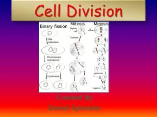
MITOSIS • Mitosis is the division of plant or animal cells in order to enable an organism to grow or repair damaged parts of itself. • Mitosis is the cell division which occurs in all body cells except for gamete (sex cell) formation. Mitosis results in the formation of two genetically identical cells, each containing the same number of chromosomes as the parent cell (the diploid or 2n number).
Mitosis ensures that the species number of chromosomes is maintained. • Mitosis ensures that each daughter cell receives an identical combination of genes.
Mitosis is the method by which all cells of a multi-cellular organism are formed from the zygote, and therefore is essential for growth. • Mitosis is the method by which organisms reproduce asexually forming offspring identical to the parent.
CENTRIOLOES AND MITOSIS • These are organelles found in the cytoplasm close to the nuclear envelope in animal and simpler plant cells. • Approximately 500nm long and 200 nm wide.
Composed of 9 groups of microtubules arranged in triplets. • Neighboring triplets are attached to each other by fibrils. • Microtubules are long hollow tubes 25nm in diameter and made of the protein tubulin.
The centrioles lie in a poorly defined area called the cemtrosome. • This area actually makes the spindle fibres. • Spindle fibres shorten by the removal of tubulin to pull chromatids apart.
The addition of the chemical colchicine to actively dividing cells inhibits spindle formation and the chromatid pairs remain in their metaphase positions. • What do you think may be an advantage to scientists of adding colchicine to dividing cells?
ANSWER • This technique enables the number and structure of chromosomes to be examined under the microscope.
The sequence of events which occurs between one cell division and the next is called the cell cycle. • It has three main stages:
1 INTERPHASE • This is a period of synthesis and growth. • The cell produces many materials required for its own growth and for carrying out all its functions. • DNA replication occurs during interphase.