
SKELETAL SYSTEM
Explore the key functions of bones in the skeletal system, including support, protection, movement, storage, and blood cell formation. Discover bone markings and fractures, learn about bone growth and development, and understand skeletal changes throughout life.
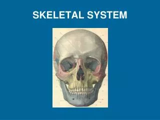

SKELETAL SYSTEM
E N D
Presentation Transcript
Functions of Bones • Support – hard framework that supports and anchors body – bones of legs act as pillars to • Protection – fused bones of the skull provide a snug enclosure for the brain – vertebrae surround the spinal cord – rib cage protects vital organs • Movement – skeletal muscles attach to bones by tendons and act as levers to move the body • Storage – bone matrix (calcium, phosphorus, potassium, sodium, sulfur, magnesium, & copper) – deposits and withdrawals are constant • Blood cell formation – hematopoiesis occurs within the marrow cavities of certain bones
Bone Markings • Projections • Tuberosity – large, rounded projection that may be roughened • Crest – Narrow ridge of bone • Trochanter – Very large, blunt, irregularly shaped process (only on the femur) • Spine – sharp, slender, often pointed projection • Process – any bony prominence
Bone Markings (ctd.) • Projections That Help Form Joints • Facet – smooth, nearly flat, articular surface • Condyle – rounded articular projection • Ramus– Arm-like bar of bone
Bone Markings (ctd.) • Depressions and Openings • Meatus – canal-like passageway • Sinus – Cavity within a bone filled with air and lined with mucous membrane • Fossa – Shallow, basin-like depression in a bone often serving as an articular surface • Fissure – Narrow, slit-like opening • Foramen – Round or oval opening through a bone
Bone Fractures, Development, and Degeneration http://www.youtube.com/watch?v=Mz2UuhbtrwY
Bone Fractures • Fracture—break in a bone • Types of bone fractures • Closed (simple) fracture—break that does not penetrate the skin • Open (compound) fracture—broken bone penetrates through the skin • Bone fractures are treated by reduction and immobilization
Common Types of Fractures Table 5.2
Hematoma Hematomaformation Stages in the Healing of a Bone Fracture Step 1: Hematoma (blood-filled swelling) is formed Figure 5.5, step 1
Hematoma Externalcallus Internalcallus(fibroustissue andcartilage) Newbloodvessels Spongybonetrabecula Hematomaformation Fibrocartilagecallus formation Stages in the Healing of a Bone Fracture Step 2: Break is splinted by fibrocartilage to form a callus Figure 5.5, step 2
Hematoma Externalcallus Bonycallus ofspongybone Internalcallus(fibroustissue andcartilage) Newbloodvessels Spongybonetrabecula Hematomaformation Fibrocartilagecallus formation Bony callusformation Stages in the Healing of a Bone Fracture Step 3: Fibrocartilage callus is replaced by a bony callus Figure 5.5, step 3
Hematoma Externalcallus Bonycallus ofspongybone Internalcallus(fibroustissue andcartilage) Newbloodvessels Healedfracture Spongybonetrabecula Bone remodeling Hematomaformation Fibrocartilagecallus formation Bony callusformation Stages in the Healing of a Bone Fracture Step 4: Bony callus is remodeled to form a permanent patch (spongy bone to compact bone) Figure 5.5, step 4
Skeletal Changes Throughout Life • Ossification Centers in a 12-week-old Fetus
Skeletal Changes Throughout Life • Fetus • Long bones are formed of hyaline cartilage • Flat bones begin as fibrous membranes • Flat and long bone models are converted to bone • Birth • Fontanels remain until around age 2
Bone Growth (Ossification) • Bones are remodeled and lengthened until growth stops • Bones are remodeled in response to two factors • Blood calcium levels • Pull of gravity and muscles on the skeleton • Bones grow in width (called appositional growth)
Articularcartilage Hyalinecartilage Spongybone New center ofbone growth New boneforming Epiphysealplatecartilage Growthin bonewidth Medullarycavity Bone startingto replacecartilage Bloodvessels Growthin bonelength New boneforming Bone collar Epiphysealplate cartilage Hyalinecartilagemodel In an embryo In a fetus In a child (a) Long Bone Formation and Growth Figure 5.4a
Long Bone Formation and Growth Figure 5.4b
The Fetal Skull • The fetal skull is large compared to the infant’s total body length • Fontanels—fibrous membranes connecting the cranial bones • Allow the brain to grow • Convert to bone within 24 months (2 years) after birth
The Fetal Skull Figure 5.13a
The Fetal Skull Figure 5.13b
Skeletal Changes Throughout Life • Adolescence • Epiphyseal plates become ossified and long bone growth ends. These plates become an Epiphyseal lineand can be seen on an X-ray. • Size of cranium in relationship to body • 2 years old—skull is larger in proportion to the body compared to that of an adult • 8 or 9 years old—skull is near adult size and proportion • Between ages 6 and 11, the face grows out from the skull
Skeletal Changes Throughout Life Figure 5.33a
Skeletal Changes Throughout Life • Osteoporosis • Bone-thinning disease afflicting • 50% of women over age 65 • 20% of men over age 70 • Disease makes bones fragile and bones can easily fracture • Vertebral collapse results in kyphosis (also known as Dowager’s Hump) • Estrogen aids in health and normal density of a female skeleton


