Acute lymphobl a stic leukemia (ALL)
310 likes | 630 Vues
Acute lymphobl a stic leukemia (ALL). Clonal proliferation and accumulation of blast cells in blood, bone marrow and other organs Disorder arises from a single lymphoid progenitor cell that has undergone genetic damage leading to dysregulated growth and arrested differentation

Acute lymphobl a stic leukemia (ALL)
E N D
Presentation Transcript
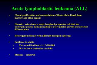
Acute lymphoblastic leukemia (ALL) Clonal proliferation and accumulation of blast cells in blood, bone marrow and other organs Disorder arises from a single lymphoid progenitorcell that has undergone genetic damage leading to dysregulated growth and arrested differentation Heterogenous disease with different biological subtypes Incidence in adults : The overall incidence 1-1,5/100 000 20% of acute leukemias in adults Etiology - unknown
Acute leukemias - clinical features 1. Bleeding 2. Fever/infection 3. Bone/joint pain 4. Hepatomegaly 5. Splenomegaly 6. Lymphadenopathy 7. CNS involvement
Symptoms and signs Patients (%) Infection/fever36 Hemorrhages 33 Lyphadenopathy57 Splenomegaly56 Hepatomegaly47 Mediastinum mass 14 CNS infiltration 7 Other organ involvement 9 Pleura2.9 Bone1.2 Pericardium 1.0 Retina 1.0 Skin0.6 Tonsils0.6 Lung0.5 Kidney0.4 Testes0.3
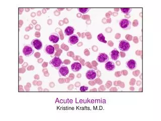
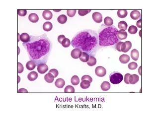
Acute leukemias - laboratory findings (1) 1. Blood examination - anemia - thrombocytopenia - variable leukocyte count, usually increased - blood morphology: presence of blast cells 2. Bone marrow morphology - presence of blast cells - suppression of normal hematopoiesis
Laboratory findings in patients with ALL at diagnosis Neutrophils (×106/L) Patients (%) <500 23 500-1000 14 1000-1500 9 >1500 54 Platelet (X 106/L) <25,000 30 25,000-50,000 22 50,000-150,000 33 >150,000 15 Hemoglobin (g/dL) <6 8 6-8 20 8-10 27 10-12 24 >12 21
Laboratory findings in patients with ALL at diagnosis Patients (%)Leukocytes (×106/L) <5,000 27 5000-10,000 14 10,000-50,000 31 50,000-100,000 12 >100,000 16 Lymphoblastsin blood smear present92 absent 8 Limphoblasts in bone marrow smear <50% 3 51%-90% 51 >90% 46 „empty” bone marrow aspiraation 16
Acute leukemias - Laboratory findings (2) 3. Cytochemical stains 4. Immunophenotyping 5. Cytogenetics 6. Molecular studies
Morphologic subtypes of acute lymphoblastic leukemias (FAB classification) Subtype Morphology Occurrence (%) L1 Small round blasts 75 clumped chromatin L2 Pleomorphic larger blasts20 clefted nuclei, fine chromatin L3 Large blasts, nucleoli,5 vacuolated cytoplasm
Acute lymphoblastic leukemias - reactivity with special stains Subtype Peroxidase or Non-specific Periodic Sudan black esterase acid-Schiff L1 - - +++ L2 - - +++ L3 - - +++
Immunologic classification of acute lymphoblastic leukemias B- lineage (80%) Markers Pro-B CD19(+),Tdt(+),CD10(-),CyIg(-), Common CD19(+),Tdt(+),CD10(+),CyIg(-), Pre-B CD19(+),Tdt(+),CD10(+),CyIg(+),SmIg(-) Mature-B CD19(+),Tdt(+),CD10(±),CyIg(±),SmIg(+) T-lineage (20%) Early-T cCD3(+) CD7(+), CD2(+/-), Tdt(+), Cortical-T cCD3(+) CD7(+), CD2(+), CD1a(+) CD4(+) CD8(+)Tdt(+) Mature-T sCD3(+) CD1a(-)
Chromosomal/molecular abnormalities with prognostic significance in ALL Better prognosis - normal koryotype - hyperdiploidy Poor prognosis - t (8; 14) - t (4; 11) Very poor prognosis - t (9; 22); BCR/ABL (+)
Risk classification in ALL 1. Standard risk 2. High risk 3. Very high risk
High-risk ALL 1. Pre – T (cCD3+/CD7+/CD2+) 2. Pro – B (CD19+/TdT+/CD10-/cIgG-) 3. Age > 35 years, 4. -WBC > 30 G/L in B-ALL > 100 G/L in T-ALL 5. No remission after 4 weeks of induction therapy 6. Detection of MRD (minimal residual disease) with flow cytometry or molecular methods- MRD positivity 7. t(4;11)
Very high-risk ALL Chromosome Philadelphia - positive or BCR/ABL (+)
Treatment phases in ALL • Remission induction therapy • Post-remission treatment • Intesification (consolidation) therapy • Haematopoietic stem cell transplantation • Maintance chemotherapy • Prophylaxis / treatment of CNS involvement • Treatment of refractory/relapsed ALL
The choice of treatment-strategy depends on: 1. Risk stratification 2. Immunophenotype of leukemic cells - T lineage, - early B lineage, - mature B lineage, 3.Age and biological condition 4. Goal of treatment
Remission induction therapy in ALL • Antineoplastic treatment • Drugs: prednisone, vincristine, antracycline, asparginase, cytosine arabinoside, cyclophosphamide • ImatinibincombinationwithchemotherapyinPh+ ALL • Treatment duration: 4-8 weeks 2. CNS prophylaxis: Mtxit, Ara-C it, steoridsit 3. Supportive care 4. Treatment of complications
The objective of induction chemotherapy = complete remission Definition of CR: • Normocellularbonemarrowwith 5% orfewerblasts • Peripheralbloodwithoutblasts • Granulocytecount > 1,0 G/L, plateletcount > 100G/L • Theabsence of anysigns and symptoms od extramedulary leukemia Complete remission: 80-90 % of pts
Intensification therapy • 8-12 weeks of chemotherapy • high dose of (HD) cytosine arabinoside, HD methotrexate, HD cyclophosphamide, asparaginase, steroids • CNS prophylaxis: Mtx it, Ara-C it, steroids it • alternatively: radiotherapy 18 Gy
Post-remission therapy in standard-risk ALL 1. Chemotherapy a/. Maintenance therapy: 6-mercaptopurine,methotrexate - for 2-3 years. b/. Intensification treatment periodically repeated: daunorubicin/adriablastin, prednisone, vincristine, cyclophosphamide. 2. CNS prophylaxis
Post-remission therapy in standard-risk ALL 1. Chemotherapy a/. Maintenance therapy: 6-mercaptopurine, methotrexate - for 2-3 years. b/. Intensification treatment periodically repeated: daunorubicin/adriablastin, prednisone, vincristine, cyclophosphamide 2. CNS prophylaxis
Post-remission therapy in high-risk ALL 1. Allogeneic hematopoietic stem cell transplantation - high-dose therapy - reduced intensity conditioning 2. Autologous HSCT (donor-) Conditioning regimen: TBI 12-13 Gy plus cyclophosphamide 120 mg/kg
Post-remission therapy in very high-risk ALL - High-dose therapy( reduced-intensity) + allogeneic stem cell transplantation Maintance therapy after alloHSCT: imatinib
Treatment results in ALL • Adults • Complete remission (CR) 85-95% • Leukemia-free survival (LFS) 40-60% • Children • Complete remission (CR) 95-99% • Leukemia-free survival (LFS) 70-80%
Treatment results LFS • Chemotherapy 30-40% • Auto-HSCT 40-45% • Allo-HSCT 45-60%