Download
1 / 23
230 likes | 259 Vues
Learn about Premature Chromosome Condensation (PCC) techniques in biological dosimetry, including methods for analyzing chromosomal aberrations post-irradiation and dose estimation issues. Explore applications of PCC in biodosimetry and mitotic fusion techniques for detecting radiation exposure. Gain insights into scoring criteria and chemical induction techniques for chromosome analysis.

E N D
Premature Chromosome Condensation (PCC) Analysis Lecture Module 6
Introduction • Biological dosimetry is generally performed by analyzing dicentrics and/or translocations at first mitosis following in vitro PHA blastic transformation • Conventional dicentric assay may not provide accurate dose estimate in case of accidental exposure above 6 Gy due to lymphopaenia, mitotic delay and cell death during culture
Lymphopaenia Mitotic delay Cell death Following exposure to high doses • At high doses, number of lymphocytes in blood is reduced and cell cycle is delayed. Therefore sometimes it is not possible to make chromosome preparations containing sufficient number of metaphase cells necessary for dose estimation by routine dicentric assay in cytogenetic dosimetry
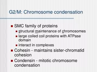
Premature Chromosome Condensation (1) PCC is research tool to probe immediate post-irradiation processes and kinetics of chromosomal break restitution and/or misrepair to form aberrations Dicentrics, complete and incomplete translocations and acentric fragments observed at metaphase are formed in G0 at differing times, dependent on dose
Premature Chromosome Condensation (2) In X- irradiated human lymphocytes: • at low doses (1-2 Gy), dicentrics and translocations are formed rapidly • at higher doses (4-6 Gy), frequencies of chromosome exchanges increase proportionally to restitution of chromosome breaks (repair)
Premature Chromosome Condensation: Techniques (1) • Techniques that cause chromatin condensation before first mitosis are termed premature chromosome condensation (PCC) assays
Premature Chromosome Condensation: Techniques (2) Premature condensation can be induced by: • Fusing interphase cells to mitotic Chinese hamster ovary (CHO) or HeLa cells using Sendai virus or polyethylene glycol (PEG) as the fusing agent • Chemical methods, using inhibitors of DNA phosphorylation such as okadaic acid or calyculin A. Most of these methods require cells to be cycling in culture
Premature Chromosome Condensation: Techniques – Mitotic Fusion • Fusing lymphocytes with CHO mitotic cells in presence of PEG, enables chromosomal aberrations to be seen immediately following irradiation without need for mitogen stimulation or culturing • This PCC method, in combination with C banding or FISH with chromosome specific DNA libraries with or without pan-centromeric probe, permits detection of breaks, dicentrics and rings as well as translocations
Techniques of Mitotic Fusion Applications in biodosimetry • useful to determine exposure to low doses and life threatening high acute doses of low and high LET radiation • discriminate between total and partial body exposures • efficient for detecting even small spared fraction (as low as 5%) • able to quantify small localized burns from partial body exposures
Mitotic fusion: Cell culture and cell fusion conditions • CHO mitotic cells from stock lines • Cells can be prepared in large quantities in advance and kept frozen before use • Isolated lymphocytes using Ficoll Hypaque, some can be frozen for future use • Use PEG as fusing agent, at 40%-50% w/v • Fuse lymphocytes with mitotic CHO cells (ratio 5:1) in presence of PEG • Fusion process takes 4 min, followed by 1h incubation in complete medium with Colcemid at 37ºC • Fixation and slide preparations by standard techniques
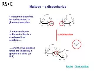
Mitotic fusion: Choose the staining technique according to the end point • PCC fragments, Giemsa (1) • dicentrics, C banding (2) • translocations, FISH (3) 1 2 3
Mitotic Fusion - Analysis • PCC spreads can be located manually or by automated metaphase finder systems • Analysis involves counting number of chromosome elements (single chromatids) • Number in excess of 46 indicates fragments • Dicentrics can be highlighted • With FISH assay, cot-1 hamster DNA can be used to mask all signals in CHO chromosomes so that only appropriate human PCC are highlighted
Mitotic fusion - Scoring criteria • Appearance of PCC defines cell cycle position of lymphocyte at time of its treatment • G0/G1 appear as single chromatid; S, as pulverized chromosomes and G2 has two chromatids • For biological dosimetry only spreads comprising single chromatids G0/G1are scored
Techniques: Chemical inductionRapid Interphase Chromosome Assay (RICA) • Isolated lymphocytes are placed in culture medium containing phosphatase inhibitor (okadaic acid or calyculin A), adenosine triphosphate and p34cdc2/cyclin B kinase and incubated at 37ºC for 3 h • Fixation and spread preparation follows normal procedures for metaphases • Radiation induced damage is analyzed after in situ hybridization and chromosome painting by fluorescence microscopy
Techniques - Chemical inductionPCC ring assay (1) • Simple and useful procedure: involves scoring of rings in Giemsa-stained chromosomes • Requires lymphocytes to be cultured • Particularly applicable to high overdoses in range where dose response for conventional dicentric assay shows signs of saturation
Techniques - Chemical inductionPCC ring assay (2) • Chemicals: okadaic acid (OA) or calyculin A is dissolved in dimethylsulphoxide (DMSO), diluted with medium and stored at -20°C as stock solution (5–10 M) • Calyculin A is about 20 times more effective than OA • Culture: PCC induction in lymphocytes requires cells to be cycling • Procedure is to PHA stimulate and culture cells for 48 h • PCC can be induced in whole blood cultures or using isolating lymphocytes
Technique – Chemical inductionPCC ring assay: Protocol • Okadaic acid (500 nM) or calyculin A (20–50 nM) is added to cultures during final 1-2 h, producing mixture of PCC cells in all stages of first cell cycle • Concentration and period of optimal treatment should be determined based on yield of PCC cells and morphology quality of chromosomes • Fixation, slide preparation and Giemsa staining procedures are similar to methods used for metaphases
Techniques – Chemical inductionPCC ring assay: Scoring criteria (1) • Select for analysis PCC cells with 46 or more chromosomal objects • In 48 h cultures of highly irradiated lymphocytes most PCC cells are between late G2 and metaphase • With low-dose exposures there may be contamination with cells at anaphase • Frequencies of PCC rings are not significantly different between late G2 and anaphase cells therefore these data can be pooled
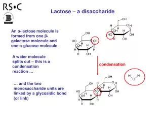
2. M/A-PCC cell with 1 ring 1. G2-PCC cell with 2 rings Techniques – Chemical inductionPCC ring assay: Scoring criteria (2)
Techniques – Chemical inductionPCC ring assay: Scoring criteria (3) • Ring with visible hole with or without centromere and pair of rings either separate or joined by centromere in PCC spreads is scored as single ring • Analyze minimum of 100 PCC-r or 500 PCC-cells • Similar to dicentric assay, overdispersed intercellular distribution of PCC rings compared with Poisson can indicate heterogeneous exposure to low LET radiation
Discussion • Drug induced PCC assay takes 48 h culturing + 2h for processing and analysis • In the dose range is 0-6 Gy conventional dicentric assay may be better dose indicator than PCC assay, but is less suitable for higher dose exposures requiring rapid dose estimate • Cell fusion induced PCC assay was not used in radiation accident biodosimetry due to highly demanding technical skill and poor yield
Conclusions Frequency of radiation induced rings and fragments in Giemsa stained PCC preparations provides useful biodosimetry tool to evaluate acute whole/partial body exposures in high dose radiation emergencies compared to conventional dicentric method PCC assay is: easy to implement amenable to automation only requires conventional light microscope cost-effective 23