Download
1 / 40
410 likes | 693 Vues
The Biomechanincs of Pitching. Tanner Neuberger. Shoulder Anatomy. Most mobile joint in the body Ball-and-socket joint 3 bones connected by muscles, ligaments and tendons Clavicle Humerus Scapula. Range of Motion. 6 degrees of freedom Flexion Elevation in the sagittal plane

E N D
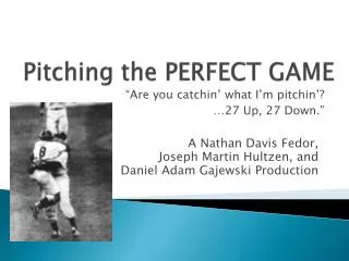
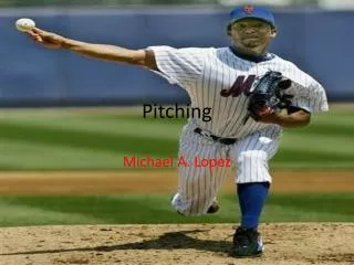
The Biomechanincs of Pitching Tanner Neuberger
Shoulder Anatomy • Most mobile joint in the body • Ball-and-socket joint • 3 bones connected by muscles, ligaments and tendons • Clavicle • Humerus • Scapula
Range of Motion • 6 degrees of freedom • Flexion • Elevation in the sagittal plane • Abduction • Elevation in the coronal plane • Rotation • Internal/external: Forearm moving in transverse plane
3 Articualtions • Acromioclavicular • Glenohumoral • Scapulothoracic AC: Restrains both rotation and posterior translation of clavicle Cocroco-acromial, coroco-clavicular, and acromio-clavicular ligaments
Sternoclavicular • Links upper extremity directly to thorax • Fibrocartilage disc between the sternum and the clavicle • Shock absorber • Held together by strong ligaments
Glenohumoral • 4 glenohumoral ligaments, connect humerus to the glenoid • Superior, middle, inferior and posterior • Inferior glenohumoral ligament splits into a front and back half • These act like a hammock to support the humoral head • Dislocation inferiorly and anterior most common in young people
Scapulothoracic • Bone-muscle-bone articulation between the scapula and thorax • Involves gliding of the scapula on the posterior aspect of the thorax • The subscapularis and the serratus anterior glide along one another • No osseous connection with the axial skeleton • Allows for a wide range of scapular motion, including protraction, retraction, elevation, depression, and rotation
Subacromial Bursa • Fluid filled sac that decreases the friction between bone and tendon • Bursa covers the rotator cuff tendons and protects them from the overlying acromion • Shock absorber
Labrum • A ring of cartilage surrounding the shoulder joint socket • Acts like a curved bumper to increase the depth of the glenoidfossa • Keeps the humeral head in the glenoidfossa and helps to prevent dislocations • Adds depth • “Break stop” or centers humeral head
Rotator Cuff • Four small muscles • subscapularis, supraspinatus, infraspinatus, and teres minor • Responsible for the stability of the shoulder joint • Holds the humeral head in the glenoid socket during early abduction
Throwing Motion • Kinetic chain concept • Sequence of body segment motions • Legs and trunk act as force generation • Shoulder is force regulation • Arm is force delivery
Phase 1: Wind-Up • Preparation of kinetic chain • Lead foot off ground • Building potential energy package • Raises center of gravity • Minimal stress to shoulder
Phase 2: Early Cocking • Positions arm in 90° abduction • Arm posterior to plane of body • Initiates external rotation • Stride initiates
Phase 3: Late Cocking • Foot contact • Maximum abduction, external rotation (40° to 170°) • Trapezius and Serratus Anterior force couple stabilizes scapula • Peak Rotator Cuff activity • Deltoid and supraspinatus function together to elevate humerus to greater than 90° of abduction
Late Cocking Cont’d • Flexion to 90° • Flexors and extensors work in coordination to control motion • Moderate valgus force exists • Problems • Anterior instability leading to internal impingement • Hyperangulation • Scapula Dyskinesis
Phase 4: Acceleration • Humerus IR 100 deg/0.5 sec • Rotates shoulder to ball release point of 90° rotation • Velocity near 7000 deg/sec • Eccentric to concentric conversion
Phase 5: Deceleration • Most violent • Ball release to 0° rotation • Eccentric contraction to slow arm • Posterior capsule stress • Joint loads • Posterior shear= 400N • Inferior shear= 300N • Compressive > 1000N
Phase 6: Follow-Through • Ball release, adduction, internal rotation, deceleration • Rebalancing • Muscles to resting level • Timing of Phases • Total 2 seconds • Wind-Up to late cocking 1.5 sec • Acceleration 0.5 sec • Deceleration to end 0.5 sec
Common Shoulder Injuries • Dislocation • Subluxation • Impingement • Rotator Cuff Tear • Dead Arm
Clavicle • Acts as a strut connecting the thorax to the upper extremity • Protects underlying brachial plexus and vascular structures • Attachment site for many of the muscles that act on the shoulder
Humerus • The head of the humerus has 2 projections, the greater and lesser tuberosities • It is at these points that the rotator cuff tendons attach
Hyperangulation • The abduction- external rotation position of the cocked baseball shoulder places a physiologic compression on the posterior labrum and posterior rotator cuff • Leads to the common problem of internal impingement • Cause • Muscular weakness (loss of dynamic stabilization) • Anterior Capsular stretch
Scapula Dyskineses • Lack of full retraction with cocking • Scapula receives load from the trunk and transfers them to the arm • Must retract and protract around thoracic wall for cocking/acceleration to deceleration • Association with tight pec major and minor, weak trapezius, serratus anterior, and rhomboids
Dislocation Cont’d • Shoulder ball and socket joint twisted apart • Occurs after a significant injury in young, active people usually under 30 years old • Older patients: accompanies other injuries such as fractures or rotator cuff tears • Subluxation occurs if the head only partially slips out and then back in • 3 major groups • Traumatic • Atraumatic • Habitual
Traumatic (anterior) Instability • Sporting injuries, major accidents/falls are the most frequent causes • Associated with structural abnormalities such as Bankart Lesion or a Hill-Sachs defect
Atraumatic Instability • Caused by repeated micro trauma to the shoulder as seen in throwing athletes • Associated with structural abnormalities such as articular surfaces damage, capsular laxity and rarely a Blankart lesion • Arthroscopic examination of the shoulder is invaluable • Treatment in 2 stages: • Physical Therapy and Injections • Surgery
Habitual Instability • Caused by inappropriate action or balance between various shoulder muscles • Diagnosed by arthroscopic examination • Treatment • Physical Therapy
Impingement • Results from abnormal contact between the greater tuberosity and the under surface of the acromion during shoulder abduction • Classically this contact occurs at a 60°-120° of shoulder abduction resulting in a painful arc in mid abduction
Cause Symptoms • Pain and weakness during activity, especially while elevating the shoulder sideways • Pain localized around the deltoid muscle and may interrupt sleep • Rotator cuff dysfunction (weakness) • Due to the degenerative changes within the rotator cuff muscles and is an age related phenomenon • May follow a painful injury or traumatic tear
Treatment • Dependant on the severity of the symptoms, age and occupation of the patient as well as whether there is an associated rotator cuff tear • If NOT associated with rotator cuff • Steroid injection into the subacromial bursa and physical therapy • If associated with rotator cuff or injections fail • Arthroscopic subacromial decompression • 90% success rate • Surgery • Rotator cuff repair may be necessary
Rotator Cuff Tear • Rotator cuff muscles are known to undergo degenerative changes with age • In some cases this could lead to pain, weakness or instability necessitating treatment
Treatment • Steroid injections, pills and physical therapy • Surgical repair of the rotator cuff in combination with subacromial decompression is best long-term outcome • Arthroscopic or open surgical techniques • Depending on the size of the tear • Postoperative recovery period is rather prolonged and may take 3-6 months • http://www.youtube.com/watch?v=IXDHvudAVOc
Dead Arm • Shoulder Stiffness due to thickening and fibrosis of the capsule • Posterior capsular repetitive micro-trauma during the follow-through phase of throwing • Lack of glenohumoral internal rotation
Treatment • Steroid and local anesthetic in glenohumoral joint and physical therapy • For severe stiffness the most effective is manipulation under general anesthetic with a steroid and local anesthetic injection into the joint and intensive physical therapy
Hill-Sachs Defect • Depression fracture caused by contact between humeral head hitting the glenoid rim • Treatment • Reduction under sedation followed by 4 weeks of immobilization in an external rotation brace
Bankart Lesion • Detachment of the cartilaginous edge of the glenoid (shoulder socket) • Creates a pocket, which in the position of shoulder abduction and external rotation allows abnormal displacement of the humeral head on the glenoid