Download
1 / 25
270 likes | 539 Vues
What are the functions of the heart?. Functions of the Heart. Did you know? At rest, 2 ounces of blood is circulated with each heart beat. It’s a PUMP. What do pumps do? How does this relate to heart function?. Heart Facts. Adult heart is about 5 inches long and 3.5 inches wide

E N D
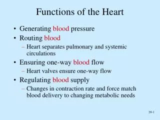
Functions of the Heart Did you know? At rest, 2 ounces of blood is circulated with each heart beat.
It’s a PUMP What do pumps do? How does this relate to heart function?
Heart Facts • Adult heart is about 5 inches long and 3.5 inches wide • Weighs < 1 pound (12 – 13 oz) • Purpose is to circulate life–sustaining blood throughout the body • Heart stops beating = life stops! • If blood flow to the brain ceases for 5 seconds or more, loss of consciousness • After 15 – 20 seconds without blood flow to the brain, the muscles twitch convulsively • After 4 – 5 minutes without blood flow brain cells are irreversibly damaged
Hear the beat! What makes the lubbdupp sound? APEX
The apex of the heart lies on the diaphragm • The apex points to the left of the body • The apex is the location where the heartbeat is most easily felt and heard through a stethoscope • To listen place the stethoscope head over the area between the 5th and 6th ribs and along an imaginary line extending from the middle of the left clavicle • During the cardiac cycle the heart valves make a sound when they close, referred to as lubbduppsounds • The lubb sound is heard first, made by the tricuspid and bicuspid closing between the atria and ventricles, heard loudest at the apex (aka the S1 sound) • The dupp sound is heard second, is shorter, higher pitched, made by the semilunar valves in the aorta and the pulmonary artery closing (aka the S2 sound) http://www.youtube.com/watch?v=oF3J4eHCU1I&feature=player_embedded
Blood Pressure Facts • Blood pressure is the surge of blood when heart pumps creating pressure against the walls of the arteries • SYSTOLIC PRESSURE • Measured during the contraction phase • Upper arm adult 120mm/Hg • DIASTOLIC PRESSURE • Measured when the ventricles are relaxed • Upper arm adult 80mmHg
It’s a PUMP What makes the pump work? It’s electric!
A heart removed from the body will continue to beat rhythmically • Showing that the heartbeat generates in the heart muscle itself • Myocardium contracts rhythmically to perform its duty as a forceful pump • Conducting cells located at the opening of the SVC into the atrium control the contractions of the heart muscle
They are known as the SA (sinoatrial node) or pacemaker • SA node sends out an electrical impulse that begins and regulates the heart • The impulse spreads out over the atria, they contract (depolarize) • Causes blood to flow down into the relaxed ventricle • The impulse reaches the AV (atrioventricular node) located between atria and ventricle
From the AV node the impulse moves to the fibers of the septum • These are called the atrioventricular bundle or the bundle of His • The bundle of His branch right and left, now called Purkinjie fibers and spread throughout the ventricles
The electrical impulse scoots along the Purkinjie fibers, causes the ventricles to contract • Then the heart rests briefly (repolarizes) • The combined actions of the SA and AV nodes make up the cardiaccycle and represents one heart beat
Each cardiac cycle takes 0.8 seconds • Average adult heart rate is 72 – 80 bpm (beats per minute) • The human heart can create enough pressure to spurt blood 30 feet!!
Electrocardiogram • aka EKG or ECG • Records the electrical activity of the heart that causes the contraction and relaxation of the atria and ventricles • SYSTOLE = contraction phase • DIASTOLE = relaxation phase • Baseline of an EKG is a flat line that separates the various waves ( P, QRS and T) • The flat line is present when there is no electrical current flowing in the heart • Waves deflect up (+) or deflect down (–) • P, QRS, and T waves indicate the contraction and relaxation phases
P = Atrial contraction QRS = Ventricular contraction T = Ventricular relaxation
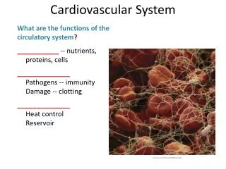
Systemic Circulation • The flow of blood after it leaves the heart and moves throughout the body and back to the heart • Circulates nutrients, O2, water and secretions to the tissues and back to heart • Carries the CO2 and other waste away from tissues • Equalizes body temp • Helps to protect the body against harmful bacteria
Cardiopulmonary Circulation • Takes deoxygenated blood to the heart to the lungs where CO2 is exchanged for O2, then returns to the heart to the body
Right side Superior vena cava Inferior vena cava Right atrium Tricuspid valve Right Ventricle Pulmonary valve Pulmonary artery Lungs CO2 and O2 exchange Left Side Pulmonary vein Left atrium Bicuspid valve Left ventricle Aortic valve Aorta Flow of blood through the Heart/Body Body
Coronary Circulation • Brings oxygenated blood to the heart muscle • The coronary artery has a right and left branch • They encircle the heart with many smaller branches to all areas of the heart muscle • Blood circulates to capillaries, gases exchange, then goes to veins • Deoxygenated blood returns through coronary veins to the coronary sinus (a trough in the posterior wall of the right atrium)
View of the Coronary sinus (trough in the posterior wall of the right atrium)
Identify the pulse sites... Dorsalispedis Temporal Brachial Radial Carotid Femoral Popliteal
Relevance of nutrients to the blood and circulatory system • The circulatory system plays a vital role in homeostasis • Absorption and transport of nutrients to cells, tissues, organs, and systems Did you know??? Vitamin K- prevents hemorrhage Vitamin B12- prevents anemia Vitamin E- prevents hemolysis