Download
1 / 48
500 likes | 838 Vues
Functions of the Heart. Functions of the Heart. Generate blood Pressure Routing Blood Ensuring one way blood flow Regulating blood supply. The Heart. Located in the Thorax Part of the Mediastinum ( heart, trachea and esophagus) Size of a Fist

E N D
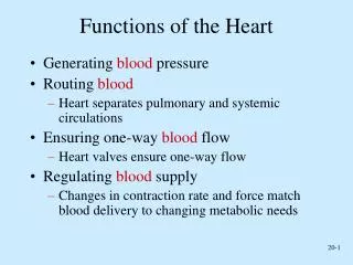
Functions of the Heart • Generate blood Pressure • Routing Blood • Ensuring one way blood flow • Regulating blood supply
The Heart • Located in the Thorax • Part of the Mediastinum ( heart, trachea and esophagus) • Size of a Fist • Blunt end is called apex, Flat portion at the opposite end is the base
The Heart • Apex- most inferior anterior and to the left located at the fifth intercostal space • Base- superior and slightly posterior most superior part is in the second intercostal space
The Pericardium • the heart is surrounded by a space called the pericardial cavity • the pericardium consists of a fibrous and a serous part • the serous pericardium consists of a parietal and a visceral pericardium
The Heart: Atria and Ventricles • Atria- located at the base • Ventricles- extends from apex to the base • Sulcus- Coronary Sulcus-extends around the heart and separates the atria from the ventricles • Vessels- veins and arteries and capillaries
Veins: • Six large veins carry blood to the heart - the superior vena cava- blood from body to right atrium - the inferior vena cava- blood from body to right atrium - four pulmonary veins- blood from lungs to the left atrium
Arteries • The pulmonary trunks and the aorta exit the heart • The pulmonary trunk arises from the right ventricle splits into right and left pulmonary arteries which carry blood to the lungs • The Aorta carries blood to body
Blood Supply to the Heart • Coronary Arteries- two coronary arteries originate from the base of the aorta just above the aortic semi lunar valves The left coronary artery originates on the left side of the aorta and it supplies most of the anterior wall of the heart and most of the wall of the right ventricle The right coronary artery originates on the right side of the aorta and supplies most of the right wall of the right ventricle
The left coronary artery originates on the left side of the aorta and it supplies most of the anterior wall of the heart and most of the wall of the right ventricle • The right coronary originates on the right side of the heart and supplies most of the wall of the right ventricle
Heart Chambers • Consists of Four Chambers Two Atria Two Ventricles
The Atria • Atria- Right and Left atria receive blood from veins Right atria- reservoir for blood returning from the body (superior vena cava, inferior vena cava and coronary sulcus) Left atria – receive the blood from four pulmonary veins
The Ventricles • The right and left ventricles of the heart are the major pumping chambers • The right ventricle pumps blood into the pulmonary arteries to the lung • The left ventricle pumps blood into the aorta which sends blood to the rest of the body
The Heart Valves • A- V ( Atrioventricular) Valves • Semilunar Valves ( Half Moon)
The A-V Heart Valves • A-V valves are located between the right atrium and right ventricle and between the left atrium and left ventricle • The A-V valves on the right are called the tricuspid valves (three cusps) • The A-V on the left side are called the bicuspid valves ( two cusps)
Heart Valves continued • Valves also exist between the right ventricle and the pulmonary arteries and the left ventricle and the aorta • These valves are called semilunar valves because they have a half moon shape.
Route of blood flow through the Right Heart • Blood returning from the body via the superior vena cava, the inferior vena cava and the coronary arteries empty into the right atrium • From the right atria blood flows through the tricuspid valve and enters the right ventricle • From the right ventricle blood flows through the pulmonary semilunar valves into the pulmonary trunk to pulmonary arteries ( to the lungs to pulmonary circulation)
Route of Blood flow through the Left Heart • Blood returning from the lungs enters the left atrium • From the left atrium blood passes through the Bicuspid valve into the left ventricle • From the left ventricle blood will then pas through the semilunar valves into the aorta and then to the systemic circulation
Animated Tutorial of the Circulation of Blood through the heart
How is the movement of the blood through the heart accomplished ? Answer?
The Cardiac Cycle To Main Pumps: • The primer pump is the atria- because they complete the filling of the ventricle • The power pump is the ventricle- because they produce the major force throught the systemic circulation
Contraction / Relaxation (Systole)/ ( Diastole) • Atrial Systole refers to contraction of the two atria • Ventricular systole refers to contraction of the two ventricle • Atrial diastole refers to relaxation of the two atria • Ventricular diastole refers to relaxation of the two ventricles
Major Events of the Cardiac Cycle Ventricular Systole- pushes blood back toward the atria, causing the A-V valves to close - pressure in the ventricles exceeds the pressure in the pulmonary trunk and aorta causing the semilunar valves to be forced open and blood is ejected into the pulmonary trunk and aorta Ventricular diastole - The semilunar valves close, preventing blood from flowing back into the ventricles . - The pressure continues to decline in the ventricle until finally the A-V valve open and the blood flows directly from the atria into the relaxed ventricle .( During the previous ventricular systole, the atria were relaxed and the AV valves open, blood flows into the ventricles and fills them to approximately 70% of their volume Atrial systole forces additional blood flow into the ventricles to complete their filling. The semilunar valves remain closed.)
Animated Tutorial of the Circulation of Blood through the heart
The Heart Sounds • A stethoscope What you can hear S1 – The first heart sound (lubb) beginning of systole results from closure of the A-v valves S2 – The second heart sound (dupp) beginning of ventricular diastole results from closure of the semilunar valves ventricular systole – between S1 and S2 ventricular diastole- between S2 and S1
Abnormal Heart Sounds continued • Murmur- result from faulty valves incompetent- the valves fail to close tightly and blood leaks backward through the valve when it is closed stenosed- the opening of the valve is narrowed usually a swishing sound
Heart Sounds Demonstrations
Regulation of Heart Function • Cardiac Output (CO)- the volume of blood pumped by either ventricle per minute • Stroke volume (SV)- is the volume of blood pumped per ventricle each time the heart contracts • Heart Rate (HR)- number of heart contractions per minute • CO= SV x HR
The Vascular System • Arteries to Capillaries to Veins
Vessel Structure • Tunica Intima • Tunica Media • Tunica Adventitia
Blood Vessels • Arteries- are blood vessels that carry blood away from the heart elastic muscular arterioles
Blood Vessels Continued • Capillaries- thin walled numerous site of gas and nutrient exchange
Blood Vessels continued • Veins- carry blood to the heart veins are thinner, with less elastic tissue venules small veins medium sized veins large veins
The Aorta • All Arteries branch from the aorta • Three parts: ascending (coronaries) aortic arch ( head and upper limbs) descending( thoracic, abdominal and common iliacs)
Blood Pressure • Blood pressure is a measure of the force blood exerts against the bllod vessel walls. • When the ventricles contract, blood is forced into the arteries , and the pressure reaches a maximum callled the systolic pressure • When the ventricles relax, blood pressure in the arteries falls to a minimum called the diatolic pressure