Impact of Cocaine Withdrawal on Brain Regions: Autoradiogram Analysis in Mice
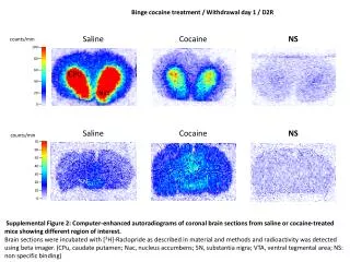
This study examines the effects of cocaine withdrawal on specific brain regions in mice, using computer-enhanced autoradiograms. The brain sections, incubated with [3H]-Raclopride, reveal differences in radioactivity between saline and cocaine-treated groups. Key regions include the caudate putamen (CPu), nucleus accumbens (Nac), substantia nigra (SN), and ventral tegmental area (VTA). Findings illustrate the alterations in dopamine receptor binding during withdrawal, enhancing our understanding of addiction and recovery processes in neuroscience.

Impact of Cocaine Withdrawal on Brain Regions: Autoradiogram Analysis in Mice
E N D
Presentation Transcript
Bingecocainetreatment / Withdrawalday 1 / D2R Saline Cocaine NS counts/min CPu 100 80 Nac 60 40 20 0 Saline Cocaine NS counts/min 70 60 50 40 SN VTA 30 20 10 0 Supplemental Figure 2: Computer-enhanced autoradiograms of coronal brain sections from saline or cocaine-treated mice showing different region of interest. Brain sections were incubated with [3H]-Raclopride as described in material and methods and radioactivity was detected using beta imager. (CPu, caudate putamen; Nac, nucleus accumbens; SN, substantianigra; VTA, ventral tegmental area; NS: non specific binding)