ChrX probes
Optimizing Hybridization Stringency for Oligonucleotide CGH Microarrays Anniek De Witte * § , Bo Curry*, Karen W. Shannon* * Agilent Technologies, Inc., 5301 Stevens Creek Blvd., Santa Clara, CA 95052 § anniek_de-witte@agilent.com.

ChrX probes
E N D
Presentation Transcript
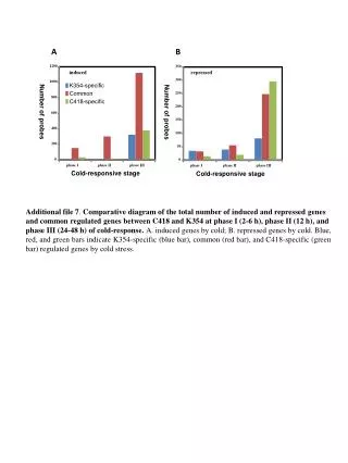
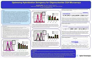
Optimizing Hybridization Stringency for Oligonucleotide CGH Microarrays Anniek De Witte*§, Bo Curry*, Karen W. Shannon* * Agilent Technologies, Inc., 5301 Stevens Creek Blvd., Santa Clara, CA 95052 §anniek_de-witte@agilent.com Detection of a simultaneous 8p deletion and 8q amplification in colorectal carcinoma cell line HT29 under optimized stringency conditions Figure 4. Log2 ratio values for all oligonucleotide probes on chromosome 8 of HT29 cell line DNA are plotted as a function of their chromosomal position using CGHAnalytics software (Agilent Technologies). Every point represents 1 probe, thin blue line shows 1 Mb moving average, thick blue lines and shaded areas show the aberration calls. Hybridizations were performed at 67°C containing 750 mM salt. The previously characterized (using BAC arrays) 8p deletion and 8q amplification and a small heterozygous loss at q23.1 were observed. Abstract Array-based comparative genomic hybridization (aCGH) is a very powerful tool used to analyze DNA copy number aberrations. Previously (1) it was shown that in situ synthesized 60-mer oligonucleotide arrays designed for CGH are able to detect chromosomal alterations throughout a genome with high sensitivity. To develop a robust platform and to obtain maximal performance, optimization of the protocol is crucial. We designed a series of experiments to determine the optimal hybridization stringency conditions for CGH microarrays. First, a male/female (XY/XX) model system was used to measure the effects of temperature, salt concentration and formamide on microarray performance. In this model system XY/XX ratios for X-chromosome probes have a theoretical value of 0.5 (log2 ratio of -1). Low-stringency hybridizations led to nonspecific binding, resulting in increased intensities and compressed log2 ratios. Relatively high-stringency hybridization conditions were found to give the best results, although increasing the stringency beyond a certain point led to lower intensities and decreased signal to noise ratios. Decreased signals were observed when a formamide-based buffer was used, possibly due to slower kinetics of hybridization at reduced temperature in the presence of formamide. Finally, analysis of a well characterized human colon tumor cell line (HT29) hybridized at the optimized stringency indicated that the data obtained with 60-mer oligonucleotide arrays agreed well with previously published BAC aCGH data. These results indicate that the optimization of stringency conditions improved microarray performance and reproducibility. (1) Barrett, M.T. et al. Proc. Natl. Acad. Sci. USA 101, 17765−17770 (2004). Effect of salt concentration on microarray performance Figure 2. The effect of salt concentration on microarray performance using a male/female (XY/XX) model system. The temperature was kept constant at 65°C. Distributions of Log2 ratio values are shown on the left panel. Lowering the salt concentration from 750 mM to 260 mM led to log2 ratios closer to the expected value indicating better separability but lowering the salt concentration too much led to low median intensities (right panel) and resulted in decreased signal to noise ratios (data not shown). 750 mM 628 mM 505 mM 260 mM ChrX probes Bin Size Autosomal probes Binned Log2Ratio Detection of a single copy gain on chromosome 18p in colorectal carcinoma cell line HT29 under optimized stringency conditions Figure 5. Log2 ratio values for all oligonucleotide probes on chromosome 18 of HT29 cell line DNA are plotted as a function of their chromosomal position using CGHAnalytics software (Agilent Technologies). Every point represents 1 probe, thin blue line shows 1 Mb moving average, thick blue lines and shaded areas show the aberration calls. Hybridizations were performed at 67°C containing 750 mM salt. The previously characterized (using BAC arrays) single copy gain on chromosome 18p could be observed. Materials and Methods Genomic DNA. Genomic DNA from normal male 46, XY and normal female 46, XX was obtained from Promega. The human colon tumor cell line HT29 was obtained from the American Type Culture Collection and was grown according to the supplier’s recommendations. Genomic DNA was isolated from this cell line using a DNeasy tissue kit (Qiagen). Oligonucleotide Microarrays. The Agilent Human Genome CGH Microarrays (44B) were used. This array consists of ~43,000 in situ synthesized 60-mer oligonucleotide probes that span coding and noncoding sequences with an average spatial resolution of ~35 kb. Labeling, Hybridization and Washing. Three micrograms of genomic DNA was digested with AluI (50 units) and RsaI (50 units) for 2h at 37°C. The digested DNA was purified with a QIAprep Miniprep kit according to the manufacturer’s protocol (Qiagen). The purified restricted samples were labeled with Cy5-dUTP or Cy3-dUTP (PerkinElmer) using the BioPrime Array CGH Genomic Labeling Module (Invitrogen). The appropriate Cy-5 labeled and Cy-3 labeled samples were combined and filtered through a Microcon YM-30 column (Millipore). The purified labeled samples were mixed with 50 μg of human Cot-1 DNA (Invitrogen) and Agilent 10x Blocking Agent (supplied with Agilent’s Oligo aCGH Hybridization Kit). In the temperature stringency experiments samples were mixed with an equal volume of Agilent 2x Hybridization Buffer. In the salt stringency experiments hybridization buffers containing different amounts of salt were used. The formamide stringency experiments were performed at a final concentration of 30% formamide. The sample was applied to the array and hybridization was carried out for 40 h at different temperatures ranging from 40°C to 70°C. The arrays were then washed in Agilent Oligo aCGH Wash Buffer 1 for 5 minutes at room temperature, followed by 1 minute at 37°C in Agilent Oligo aCGH Wash Buffer 2. Imaging and Data Analysis. Slides were scanned on an Agilent 2565BA microarray scanner. The images were analyzed by using Feature Extraction software (version 8.1, Agilent Technologies) and CGHAnalytics software (version 3.2, Agilent Technologies). Effect of formamide on microarray performance Figure 3. The effect of formamide in the hybridization buffer on microarray performance using a male/female (XY/XX) model system (F = 30% formamide, A = aqueous). Distributions of Log2 ratio values are shown on the left panel. Log2 ratios were closer to the expected value indicating better separability when the temperature was increased but aqueous hybridizations at both 65°C and 67°C (750 mM salt) showed improved log2 ratios. Additionally, in the presence of formamide some specific probes performed worse than expected. Lower signals were observed when a formamide-based buffer was used compared to an aqueous buffer, possibly due to slower kinetics of hybridization at reduced temperature (right panel). 40°C (F) 45°C (F) 50°C (F) 65°C (A) 67°C (A) ChrX probes Bin Size Autosomal probes Detection of a homozygous deletion at the A2BP1 locus in colorectal carcinoma cell line HT29 under optimized stringency conditions Figure 6. Log2 ratio values of oligonucleotide probes on a zoomed-in region of chromosome 16 of HT29 cell line DNA are plotted as a function of their chromosomal position using CGHAnalytics software (Agilent Technologies). Every point represents 1 probe with no moving averages, thick blue line and shaded area shows the aberration call. Hybridizations were performed at 67°C containing 750 mM salt. A homozygous deletion at the A2BP1 locus could be observed. Effect of hybridization temperature on microarray performance Figure 1. The effect of temperature on microarray performance using a male/female (XY/XX) model system. The salt concentration was kept constant at 750 mM. In this model system XY/XX ratios for X-chromosome probes have a theoretical value of 0.5 (log2 ratio of -1). Distributions of Log2 ratio values are shown on the left panel. Log2 ratios were closer to the expected values indicating better separability when the hybridization temperature was increased from 60°C to 70°C. Corresponding median intensities decreased with increasing temperature (right panel), indicating that low-temperature hybridization led to nonspecific binding. 60°C 62.5°C 65°C 67.5°C 70°C Binned Log2Ratio ChrX probes Bin Size Autosomal probes • Conclusions • Specificity and signal intensity must be balanced, such that the signal is maximized while minimizing non-specific probe binding. • The sensitivity of the Agilent aCGH system enables the use of higher stringency to achieve higher specificity. Optimal aCGH results are achieved with hybridizations at a final concentration of 750 mM salt and a hybridization temperature of 65°C. • Data obtained on Agilent 60-mer oligonucleotide CGH arrays with a well characterized human colon tumor cell line (HT29) hybridized at the optimized stringency agrees well with previously published BAC aCGH data. Binned Log2Ratio