Download
1 / 33
600 likes | 4k Vues
Definition of anemia. 1. Reduction in the hemoglobin concentration in blood 2. Decreased total circulating red cell mass. Normal values for peripheral blood. Female Male Erythrocytes ( per µ l ) 4.8±0.6x10 6 5.4±0.8x10 6 Hemoglobin ( g/dl ) 14 ±2 16 ±2

E N D
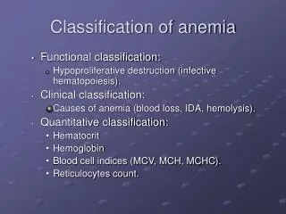
Definition of anemia 1. Reduction in the hemoglobin concentration in blood 2. Decreased total circulating red cell mass
Normal values for peripheral blood Female Male Erythrocytes(per µl) 4.8±0.6x10⁶ 5.4±0.8x10⁶ Hemoglobin (g/dl) 14 ±2 16 ±2 Hematocrit(%) 42 ±5 47 ±5 Reticulocytes(%) 1 1 ___________________________________________ Mean corpuscular volume (MCV; µm3)82-92 Mean corpuscular hemoglobin (MCH; pg)27-32 Mean corpuscular hemoglobin concentration (MCHC; %) 32-36
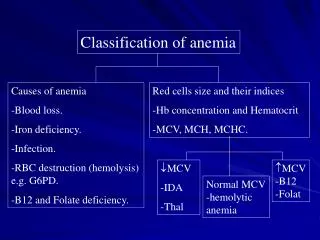
Etiologic classification of anemias • Impaired red cell production A. Disturbance of proliferation and differentiation of stem cells B. Disurbance of proliferation and maturation of erythrocytes II. Increased rate of destruction (hemolytic anemias) A. Intrinsic abnormalities B. Extrinsic abnormalities
Etiologic classification of anemias (1) I. Impaired red cell production A. Disturbance of proliferation and differentiation of stem cells ( aplastic anemia, pure red cell aplasia) B. Disurbance of proliferation and maturation of erythrocytes: 1.Defective DNA synthesis (megaloblastic anemias) 2.Defective Hb synthesis: a/. Deficient heme synthesis (iron deficiency) b/. .Deficient globin synthesis (thalassemia) 3. Unknown or multiple mechanisms (anemia of chronic disease, anemia of marrow replacement)
Etiologic classification of anemias (2) II.Increased rate of destruction (hemolytic anemias) A. Intrinsic abnormalities Hereditary 1. Red cell membrane defects (hereditary spherocytosis, hereditary eliptocytosis) 2. Red cell enzyme deficiencies a/. Glycolytic enzymes: pyruvate kinase, hexokinase b/. Enzymes of hexose monophosphate shunt: G-6PD, glutathione synthetase 3. Disorders of globin synthesis a/. Deficient globin synthesis (thalassemia) b/. Structurally abnormal globin synthesis (sickle cell anemia, unstable hemoglobins) Acquired 1. Membrane defect: paroxysmal nocturnal hemoglobinuria
Etiologic classification of anemias (3) B. Extrinsic abnormalities 1. Antibody mediated a/. Autoantibodies (idiopathic, drug-associated, SLE, malignancies) b/. Isohemagglutinins (transfusion reactions, erythroblastosis fetalis) 2. Mechanical trauma of RBC a/. Microangiopathic hemolytic anemias (thrombotic thrombocytopenic purpura, DIC) b/. Cardiac traumatic hemolytic anemia 3. Chemicals and microorganisms 4. Sequestration in mononuclear phagocytic system - hypersplenism
Classification of anemias (simplified) 1. Deficiency anemias 2. Aplastic anemia 3. Hemolytic anemias 4. Secondary anemias
Morphologic classification of anemias Type MCV MCHC Common cause ________________________________________________________ Macrocytic anemiaincreased normal Vitamin B12 deficiency Folic acid deficiency Microcytic anemia - hypochromicdecreased decreased Iron deficiency Thalassemia - normochromicdecreased normal Spherocytosis or normal Normocytic anemianormal normal Aplastic anemia - normochromicChronic renal failure Some hemolytic anemia
Stem cell disorder 1. Aplastic anemia 2. Pure red cell aplasia A. Congenital B. Acquired
APLASTIC ANEMIA (AA) • Definition • AA is characterized by pancytopenia with hypocellular marrow; hematopoietic tissue replaced by fat cells, in absence of abnormal infiltrate or increase in reticulin • Incidence (acquired) • 2/1000000 • rare < 1 year; plateaus 20-60 yrs; increase > 60 yrs
Causes of aplastic anemia (1) • Primary (idiopathic) 70-80%: immune-mediated disease II. Secondary - drugs 1. Unpredictable (idiosyncratic reaction) - antiepileptic drugs (hydantoins) - oral antidiabetic agents (tolbutamide, chlorpropamide) - tranquillizers (chlorpromazine, chlordiazepoxide) - antirheumatic drugs (gold, indomethacin, phenylobutazone) - antibacterial agents (sulfonamides, isoniazid, steptomycin, tetracyclines, chloramphenicol) 2. Unpredictable hypersensitivity (immune reaction) - many drugs
Causes of aplastic anemia (2) III. Associated diseases 1. viral hepatitis 2. CMV infection 3. EBV infection 4. Parvovirus B19 5. paroxysmal nocturnal hemoglobinuria IV. Industrial and household chemicals: benzene, some organic solvents, trinitrotoluene, certain insecticides (DDT, chlordane, lindane)
Causes of marrow aplasia 1. Ionizing radiation 2. Antineoplastic drugs: - folic acid antagonists, - alkylating agents, - anthracyclines, - nitrosoureas - purine and pyrimidine analogous
PATHOGENESIS OF AA • Quantitative or qualitative abnormalities of pluripotent stem cell • Abnormal humoral or cellular control of hematopoiesis • Abnormal hematopoietic microenvironment • Immunologic suppression of hematopoiesis
Diagnosis of aplastic anemia 1. Personal medical history; family history 2. Physical examination 3.Clinical symptoms: - infections - bleeding - symptoms of anemia 4. Laboratory findings: - anemia, neutropenia, thrombocytopenia - bone marrow: hypocellular with fatty changes
Criteria for diagnosis of AA (1) 1. Cytopenia - Hb <10g/dL - ANC <1,5 G/L - PL <100 G/L 2. Bone marrow histology and cytology - decreased marrow cellularity (< 25%) - increased fat cells component - no extensive fibrosis - no malignancy or storage disease
Criteria for diagnosis of AA (2) 3. No preceding treatment with X-ray or antyproliferative drugs 4. No lymphadenopathy or hepatosplenomegaly 5. No deficiencies or metabolic diseases 6. No evidence of extramedullary hematopoiesis
Classification of aplastic anemia 1. Severe aplastic anemia is defined if at last two of the following criteria are present: - ANC < 0.5 G/l - PLT < 20 G/l - RTC < 1% (20 G/l) Hypoplastic bone marrow (less than 25%) on biopsy 2. Very severe aplastic anemia - criteria as above but ANC < 0.2 G/l 3. Non-severe aplastic anemia.
Prognosis of SAA if supportive therapy are only applied The overall mortality is 65-75% and the median survival 3 months
Management of severe aplastic anemia 1. Hematopoietic stem cell transplantation 2. Immunosuppressive treatment - cyclosporine - antilymphocyte/antityhymocyte globulin, - methylprednisolone - growth factors (G-CSF) 3. Androgens 4. Supportive therapy 5. Growth factors (GM-CSF, G-CSF, EPO)
Hematopoietic stem cell transplatation in severe aplastic anemia 1. Advantages - correction of hematopoietic defect - long-term survival: 80% - 90% (HLA-matched sibling donor) - majority of the patients appear to be cured 2. Restrictions - age below 40 - suitable donor available in less than 30% (sibling) - 25-40% risk of GVHD - 5-15% risk of graft failure in multitransfused patients -high mortality after MUD-HSCT - solid tumors (12%)
1st Complete Remission 2nd Complete Remission Not in Remission Chronic Phase Accelerated Phase Blast Phase 100-DAY MORTALITY AFTER HLA-IDENTICAL SIBLING TRANSPLANTATION 2001-2002 100 80 60 MORTALITY, % 40 20 0 AML ALL CML MDS AplasticAnemia ImmuneDeficiency 11
1st Complete Remission 2nd Complete Remission Not in Remission Chronic Phase Accelerated Phase Blast Phase 100-DAY MORTALITY AFTER UNRELATED DONOR TRANSPLANTATION 2001-2002 100 80 60 MORTALITY, % 40 20 0 AML ALL CML MDS AplasticAnemia ImmuneDeficiency 12
Non-myeloablative (N=7532) Traditional (N=36,543) INDICATIONS FOR ALLOGENEIC BLOOD AND MARROW TRANSPLANTS REGISTERED WITH THE IBMTR, 1997-2002- Worldwide - 14,000 12,000 10,00 8,000 TRANSPLANTS 6,000 4,000 2,000 0 AML CML ALL MDS/MPSOtherLeukemia NHL MultipleMyeloma CLL HodgkinDisease RenalCell OtherCancer SAA OtherNon-MalignantDisease
Results of combined immunosuppressive therapy in severe aplastic anemia 1. Antilymphocyte/antithymocyte globulin with androgens 43% 2. Cyclosporine and androgens 45% 3. Antilymphocyte/antithymocyte globulin and methylprednisolone 47% 4. Antilymphocyte/antithymocyte globulin, methylprednisolone and cyclosporine A 60-70%
Complication of immunosuppressive therapy • Failure of therapy and relapse of AA • exhaustion of stem cell reserves • insufficient immunosuppression • misdiagnosis (MDS) • hereditary bone marrow failure (non-immune pathogenesis) 2. Hematopoietic clonal disease • acute myelogenous leukemia • myelodysplastic syndrom • paroxysmal noctutnal hemoglobinria
Novel agents in treatment of AA(immunosuppresive, immunomodulators) • Mycofenolate mofetil (Cellcept) • Anti-Il-2 receptor monoclonal antibody (daclizumab; Zenapax) • Anti CD52 monoclonal antibody (alemtuzumab; Campath -1H) • Rapamycin • Anti-TNF alfa monoclonal antibody (etanercept; Enbrel)
Therapy of non-severe aplastic anemia 1. „Watch and wait” 2. Androgens (?) 3. Supportive care: blood and platelet transfusion, antibiotics, growth factors 4. Immunosuppressive treatment in selected patients
Androgens in the treatment of AA 1. Severe aplastic anemia - no effect when applied as a single agent, - improve the results if in combination with antilymphocyte/antithymocyte globulin and cyclosporine 2. Non-severe aplastic anemia - effective in 20 - 30% of patients.
Causes of pancytopenia 1.Failure of production of blood cells a) bone marrow infiltration - acute leukemias - hairy cell leukemia - multiple myeloma - lymphoma - myelofibrosis - metastatic carcinoma b) aplastic anemia c) vit.B12 and folate deficiency 2. Ineffective hematopoesis - myelodysplastic syndrome 3. Increased destruction of blood cells - hipersplenism - autoimmune disorders - paroxysmal nocturnal hemoglobinuria 4. Myelosuppression after irradiation or antiproliferative drugs
Case report 1 • P.W. 18-years-old student • January 2002 : appendectomy • April 2002 : hepatitis B • June 2002 : progressive pancytopenia • July 2002 : SAA • September 2002 : BMT from sibling donor • alive and healthy
Case report 2 • P.S. 16-years-old girl • February 2000 : non-severe aplastic anemia • blood transfusion • November 2001 : immunosuppresive treatment • without improvement • May 2002 : BMT from sibling donor • complete recovery before +30 day • June 2002 : died because of TTP
Case report 3 • M.R. 25-years-old woman • June 2002 : severe aplastic anemia • no sibling donor • July 2002 : immunosuppresive treatment • without improvement • December 2002 : immunosuppresive treatment • improvement ( without blood and platelet transfusion) • November 2003 : relapse of SAA • September 2004 : PBSCT from unrelated donor • alive and haelthy