ANEMIA
ANEMIA . IMS 423 BLOCK. Dr. Shaikh Mujeeb Ahmed Assistant Professor AlMaarefa College. Intended Learning Outcomes. Describe pathophysiology of anemia Enumerate the different types of anemia Describe the clinical features of anemia


ANEMIA
E N D
Presentation Transcript
ANEMIA IMS 423 BLOCK Dr. ShaikhMujeeb Ahmed Assistant Professor AlMaarefa College
Intended Learning Outcomes • Describe pathophysiology of anemia • Enumerate the different types of anemia • Describe the clinical features of anemia • Interpret the lab results and correlate the findings with the type of anemia.
ANEMIA • What is Anemia? • Anemia means - Decreased hemoglobin - Decreased RBC count - Decreased Hematocrit [PCV] • Therefore, decreased O2 carrying capacity of blood.
BLOOD • Consists of 3 types of specialized cellular elements suspended in plasma (liquid portion of blood) • Erythrocytes • Red blood cells • Important in O2 transport • Leukocytes • White blood cells • Immune system’s mobile defense units • Platelets • Cell fragments • Important in hemostasis
ERYTHROCYTES • Normal RBC count - 5 million per cubic millimeter (mm3) of blood. • Male – 5 – 5.5 / mm3 • Female 4.5 – 5 / mm3 • RBC contain hemoglobin which carries O2. • Main function of RBC – O2 transport, also CO2 transport.
STRUCTURE OF RBC • RBC are biconcave discs 7.5 - 8 micrometer (µm) in diameter and 2µm thick at outer edge and 1µm thick at the center. • RBC membrane is flexible and can change as RBC pass through capillary with a narrow diameter of 5µm.
HEMOGLOBIN • Hemoglobin is found only in RBC. • Normal Hemoglobin – 15 gram / dl . • Structure of Hemoglobin • It has two parts 1.Globin – protein has 4 polypeptide chain 2 αchain [141 amino acid in each chain] 2 β chain [146 amino acid in each chain] 2.Heme – 4 iron containing groups, each is bound to one polypeptide chain.
HEMOGLOBIN[cont] • Each iron atom present in Heme [iron is in ferrous state] can combine reversibly with one molecule of O2, therefore, each hemoglobin molecule can take four O2 molecules in the lungs. • 98.5% of O2 is carried in the blood bound to hemoglobin. • Hemoglobin is a pigment naturally colored because of iron content.
HEMOGLOBIN[cont] • It appears reddish when combine with O2, e.g. Arterial blood. • It appears bluish when deoxygenated, e.g. venous blood.
HEMOGLOBIN FUNTIONS • Transports O2. • Also transports CO2. • Combines with H+ ion, therefore, plays part as buffer. • Combines with carbon monoxide (CO), therefore, can cause CO poisoning. • Nitric Oxide (NO) gas combines with hemoglobin and this NO is released at the tissues and causes vasodilation.
IMPORTANT NOTE • RBC is mainly a plasma membrane having hemoglobin. • RBC has no nucleus and organelle. • Enzyme in RBC - Glycolytic enzyme, it generates energy ATP for active transport at membrane. - Carbon anhydrase enzyme for CO2 transport.
ERYTHROPOIESIS • Formation and maturation of RBCs. • In adult RBC are formed in bone marrow. [Bone marrow is cellular tissue that fills the internal cavities of bones]. • Bone marrow normally generates new RBC to replace old ruptured cells. • In the fetus – RBC formation takes place in yolk sac during first 03 months of life then liver and spleen up to 7th month of intrauterine life. • Bone marrow starts from 4th month till birth of baby.
ERYTHROPOIESIS • In children, most bones produce RBC by red bone marrow then red bone marrow is replaced by fatty yellow bone marrow that does not produce RBC. • In adults, red bone marrow remains in sternum, ribs, vertebrae, pelvis, upper end of long bones e.g. femur, humerus.
IMPORTANT • If we need bone marrow sample for examination, we usually take from iliac crest or sternum.
ERYTHROPOIESIS • As RBC matures, it involves - reduction in size - disappearance of nucleus - acquiring of hemoglobin
NUTRIONAL REQUIREMENT OF RBC PRODUCTION • 1. Amino Acids – for synthesis of globin of hemoglobin. • 2. Iron – If iron deficiency, it causes microcytic hypochromic anemia [small RBC with less Hb]. • 3. Vitamins – Vitamin B12 and folic acid for synthesis of nucleo protein. If less DNA metabolism affected and results in megloblastic anemia [mega means large]. • 4. Trace elements – e.g. copper, zinc, cobalt • 5. Hormones – Cortisol, growth hormone.
CONTROL OF ERYTHROPOIESIS • It is done by Erythropoietin hormone. • Source of Erythropoietin – mainly kidney. • Erythropoietin is produced by the kidneys due to reduced O2 delivery to kidney. • Main stimulus for production of erythropoietin is hypoxia e.g. high altitude, anemia. • Hormone erythropoietin is secreted in blood and stimulates erythropoiesis in the bone marrow by acting on committed RBC.
IMPORTANT • Normal RBC count 5 millions / mm3. • In every person, 25 trillion – 30 trillion RBC are moving through our blood vessels. • Average life of RBC is 120 days. • RBC are replaced at average rate of 2 millions to 3 millions / sec.
RETICULOCYTES • It is immature erythrocyte. • Normal reticulocyte count 0.5 – 1.5% in blood. • Increased reticulocyte count in blood indicates high rate of erythropoietic activity.
SYNTHETIC ERYTHROPOIETIN • Synthetic erythropoietin is given to kidney failure patients or those patients under going chemotherapy for cancer as chemotherapy affects bone marrow and developing RBC.
RBC BREAKDOWN • Average life of RBC is 120 days then it is destroyed. • When RBC breakdown, they release hemoglobin. • Hemoglobin is taken by macrophages. • Hemoglobin is broken into heme + globin. • Globin is degraded into amino acids which are used. • From Hemoglobin, iron is released and passes back to blood. Porphyrin portion of hemoglobin molecule is converted into bilirubin. • Bilirubin is carried to liver [bound with albumin] and secreted in bile by liver.
WHAT YOU SHOULD KNOW FROM THIS LECTURE ? • Normal RBC count, Size, Shape and Function • Life Span of RBC • Erythropoiesis in Adults & Children • Nutritional Requirement for Erythropoiesis • Erythropoietin • Functions of Hemoglobin • Importance of Reticulocyte count in blood • Hemoglobin Breakdown
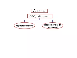
CLASSIFICATION OF ANEMIA • ON THE BASIS OF • Size of the RBCs • Normocytic • Microcytic • Macrocytic • Hemoglobin content • Normochromic • Hypochromic
CLASSIFICATION OF ANEMIA We will define MCV, MCH, MCHC. • Mean Corpuscular Volume (MCV) it is the volume of average RBC - Normal MCV = 90 fL or 90 μ3 [MCV > 95 fL are called macrocyte] [MCV < 80 fL are called microcyte] • Mean Corpuscular Hemoglobin (MCH) – it is mean concentration of Hemoglobin in each RBC. - Normal MCH = 30 picogram [pg] • Mean corpuscular Hb concentration (MCHC) – it is hemoglobin present per 100ml of RBC. - Normal MCHC = 30 gram/100ml of RBC
NORMAL VALUEs & FORMULAE FOR CALCULATION OF MCV, MCH, MCHC • Mean Corpuscular Volume (fl) • 78 – 98 (fl) • Mean Corpuscular Hemoglobin (pg) • 27 – 33 pg. • Mean corpuscular Hb concentration • 30 – 35%
CLINICAL FEATURES OF ANEMIA • Pallor • Tachycardia • Hyperbilirubinemia, Jaundice • Increase level of iron absorption – damage to endocrine glands
BLOOD LOSS ANEMIA • Rate of hemorrhage • Bleeding is internal / external • Acute loss • Loss of intravascualr volume • Cardiovascular collapse • Normocytic normochromic type • Increase reticulocyte count • External bleeding • Iron loss – iron deficiency anemia • GI bleeding, menstrual bleeding • Chronic loss - Microcytic hypochromic anemia
HEMOLYTIC ANEMIA • Characterized by; • Premature destruction of RBCs • Iron retention • Hb destruction products • Normocyticnormochromic anemia • Increase reticulocyte count • Presents with; • Fatigability • Dyspnea • Cause • Intrinsic • Cell membrane • Enzymes • Hemoglobin • Extrensic • Immune mechanism • Mechanical trauma • Infection
Inherited Disorder of the Red Cell Membrane • Heredditaryspherocytosis • Autosomal dominant triat • Abnormality of spectrin & ankyrin membrane protein • Sphere shape cells • Mild hemolytic anemia • Jaundice • Spleenomegaly • Bilirubin & • Gall stone • Treated with; • Splenectomy • Blood transfusion
Inherited Disorder of the Red Cell Membrane • Sickle cell disease • Abnormal Hb S • Chronic hemolytic anemia • Mean life span of RBCs is 20 days • Pain • Organ failure • HbS gene as recessive inheritance • Sickle cell trait • Sickle cell disease • 30% in African population;
Definition • Autosomal recessive genetic disease: • β-globin gene (chromosome 11q) mutation GAGGTG at 6thcodon • Glutamic Acid Valineat the 6th amino acid along the β-globin chain • α2β2 = normal hemoglobin • α2βS = heterozygote = Sickle trait • α2S2 = homozygous recessive = Sickle cell disease
Sickling mechanism Hb SS • Precipitating Factors • Hypoxia • Acidosis • Dehydration • Infections Deoxygenated Polymerise Pseudocrystalline Tactoids
Clinical course • Homozygous for HbS experiences • Severe hemolytic anemia • Hyperbilirubinemia • Jaundice • Gall stones • Vaso-occlusive crises
Complication • Vaso-occlusive pain crises • Acute chest syndrome
Vaso-occlusive pain crisis • Common sites • Abdomen • Chest • Bone • Joints • Infarction of • Liver • Spleen • Kidney • Heart • Retina
Acute chest syndrome • Atypical pneumonia resulting from pulmonary infarction • Pulmonary infiltrates • Shortness of breath • Fever • Chest pain • Cough
Aplastic crises • Infection with erythrovirus 19 • Severe red cell aplasia • Reduced reciculocyte count
Diagnosis & treatment • Clinical findings + blood smear – sickle cells • Hemoglobin solubility result • RBCs + sodium dithionate = turbid solution • Hemoglobin electrophoresis Management • Prophylactic treatment with penicillin • Immunization for • H. influenzae • Hepatitis B • Pneumococcal vaccine
Diagnosis & treatment • Vaso-occlusive crises • Rehydration, analgesics, antibiotics, transfusion • Hydroxyurea – to reduce pain crises • Bone marrow & stem cell transplantation • Gene therapy