ANEMIA
ANEMIA. Dr. Mansour Aljabry Assistant professor & Consultant Hematologists. Hemoglobin??. Hemoglobin structure. α. β. Globin chain. O2. O2. Fe⁺⁺. Haem. Fe⁺⁺. α. β. Prophyrin ring . Iron atom. Fe⁺⁺. Fe⁺⁺. O2. O2. Dr. Aljabry. Hemoglobin.


ANEMIA
E N D
Presentation Transcript
ANEMIA Dr. MansourAljabry Assistant professor & Consultant Hematologists
Hemoglobin structure α β Globin chain O2 O2 Fe⁺⁺ Haem Fe⁺⁺ α β Prophyrin ring Iron atom Fe⁺⁺ Fe⁺⁺ O2 O2 Dr. Aljabry
Hemoglobin • Hemoglobin is the protein molecule in RBC that carries O2 from the lungs to the body's tissues and returns carbon CO2 from the tissues back to the lungs. • Hemoglobin maintains the shape of RBC also.
Hematopoiesis Myeloid SC Erythroid Precursors Hematopoietic stem cell: 1- Self renewal 2- Cell differentiation Erythropoietin GATA1 Transcriptional Factor
Erythropoiesis The “Bone Marrow” is the major site with the need of: Folic acid – Iron “Ferrous” – Vit B12 – Erythropoietin -Amino acids minerals - other regulatory factors Basophilic Normoblast Erythroblast Intermediate Normoblast Late Normoblast Reticulocyte Erythrocyte +++ ++ + - ++ + Synthesis of Hemoglobin 6
Normal Ranges HCT Hb MCH MCV Microcytic Hypochromic Normocytic Macrocytic Normochromic
ANEMIA • An (without) -aemia (blood) • Reduction of Hb concentration below the normal range for the age and gender • Leading to decreased O2 carrying capacity of blood and thus O2 availability to tissues (hypoxia)
Clinical Features • Presence or absence of clinical feature depends on: • 1-Speed of onset: • Rapidly progressive anemia causes more symptoms than slow onset anemia due to lack of compensatory mechanisms: • (cardiovascular system, BM &O2 dissociation curve • 2-Severity: • Mild anemia :no symptoms usually • Symptoms appear if Hb less than 9g/dL • 3- Age: • Elderly tolerate anemia less than young patients
Clinical Features • Weakness • Headache • Pallor • Lethargy • Dizziness • Palpitation (tachycardia) • Angina • Cardiac failure Related to anemia Related to compensatory mechanism
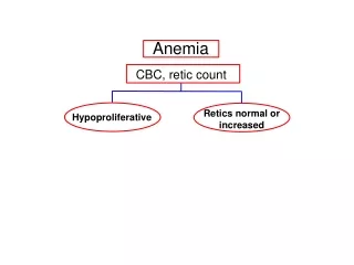
Hemoglobin RBC count DNA Prophyrin DNA synthesis Blood loss Iron Hemolysis RBC production Globin chain Acute bleeding Thalassemia Autoimmune Enzymopathy Membranopathy Mechanical Sickle cell anemia • BM failure: • -Chemotherapy • -Aplastic anemia • -Malignancy • Anemia of chronic disease Megaloblastic anemia: -B12 def. -Folate def. MDS Iron def. anemia Sidroblastic anemia Macrocytic anemia Hypochromic microcytic anemia Normocytic normochromic anemia
Iron Deficiency Anemia • Iron is among the abundant minerals on earth (6%). • Iron deficiency is the most common disorder( 24%). • Limited absorption ability : • 1-Only 5-10% of taken iron will be absorbed • 2- Inorganic iron can not be absorbed easily. • Excess loss due to hemorrhage !
Iron cycle and storage Storage forms: Ferritin Haemosiderin
IL6 Tfr2 Hypoxia +ve Hepcidin - ve Iron for erythropoeisis BM macrophage
Iron Absorption • 1-Body Iron status: Increased demands (iron def.,pregnancy..) Low iron stores high absorption Iron overload Full iron stores Low absorption 2- Content and form of dietary iron More Iron More absorption Heam Iron Ferrous Iron 3- Balance between dietary enhancers&Inhibitory factors: Enhancers Inhibitors Meat (haem iron) Fruit (Vitamin C) Sugar (Solubilizing agent ) Acids Dairy foods (calcium) High fiber foods (phytate) Coffee &tea (polyphenoles) Anti -Acids
Causes of IDA • 1-Chronic blood loss: • GIT Bleeding: peptic ulcer, esophageal varices , hookworm cancer • Uterine bleeding • Hematuria • 2- Increased demands: • Immaturity • Growth • Pregnancy • EPO therapy • 3-Malabsorption: • Enteropathy • Gastrectomy • 4-Poor diet: Rare as the only cause (rule out other causes)
Development of IDA Signs of anemia
Signs and symptoms of IDA a c b • Beside symptoms and signs of anaemia +/- bleeding patients present with: • (a): Koilonychia(spoon-shaped nails) • (b): Angular stomatitis and/or glossitis • (c): Dysphagia due to pharyngeal web (Plummer-Vinson syndrome)
Investigation normal • Microcytic hypochromic anemia with: • Anisocytosis( variation in size) • Pokiliocytosis (variation in shape)
Iron Studies Normal IDA TIBC* high TIBC • Serum Iron • Serum ferritin • Transferrin saturation • Low serum iron • Low serum ferritin • Low transferrin saturation TIBC : total iron binding capacity of transferrin
Iron Studies Thalassemia Normal IDA Low TIBC* high TIBC • High Serum Iron • High Serum ferritin • High Transferrin saturation • Low serum iron • Low serum ferritin • Low transferrin saturation
Investigation BM Iron stain (Perl’s stain): The gold standard but invasive procedure Normal IDA: reduced or absent iron stores (hemosiderin)
Treatment of IDA • Treat the underlying cause • Iron replacement therapy: • Oral :( Ferrous Sulphate OD for 6 months) • Intravenous:( Ferric sucrose OD for 6 months) Hb should rise 2g/dL every 3 weeks
PREVENTION OF IDA • Dietary modification • Meat is better source than vegetables. • Food fortification (with ferrous sulphate) • GIT disturbances ,staining of teeth & metallic taste. • Iron supplementation: • For high risk groups.
Anemia of chronic disease • Normochromic normocytic (usually) anemia caused by decreased release of iron from iron stores due to raised serum Hepcidin . • Associated with • - Chronic infection including HIV, malaria • - Chronic inflammations • -Tissue necrosis • -Malignancy
IL-6 IL-1 TNF Tuberculosis SLE Carcinoma Lymphoma +ve Hepcidin - ve no Iron for erythropoeisis BM macrophage
Work-up and treatment • Normocytic normochromic or mildly microcytic anaemia • Low serum iron and TIBC • Normal or high serum ferritin ( acute phase reactant) • High haemosiderin in macrophages but low in normoblasts Management: Treat the underlying cause Iron replacement +/- EPO