Download
1 / 2
20 likes | 164 Vues
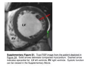
LV. RV. Supplementary Figure S1 . True-FISP image from the patient depicted in Figure 3A . Solid arrows delineate compacted myocardium. Dashed arrow indicates epicardial fat. LV left ventricle; RV right ventricle. Systolic function can be viewed in the Supplementary Movie. A. B. C.

E N D
LV RV Supplementary Figure S1.True-FISP image from the patient depicted in Figure 3A. Solid arrows delineate compacted myocardium. Dashed arrow indicates epicardial fat. LV left ventricle; RV right ventricle. Systolic function can be viewed in the Supplementary Movie.
A B C Supplementary Figure S2. Interobserver variability. A: Signal intensity, used to determine presence or absence of LGE. B: Maximum NC:C layer thickness ratio. C: Left ventricular end-diastolic volume (LVEDV).