Histology: Connective Tissue
Histology: Connective Tissue. J. Matthew Velkey matt.velkey@duke.edu 452A Davison. Connective Tissue. Epithelium. Epithelium. Connective Tissue. ridge (peg). papilla. Connective Tisue. Muscle. General Properties of Connective Tissue.

Histology: Connective Tissue
E N D
Presentation Transcript
Histology:Connective Tissue J. Matthew Velkey matt.velkey@duke.edu 452A Davison
Connective Tissue Epithelium Epithelium Connective Tissue ridge (peg) papilla Connective Tisue Muscle
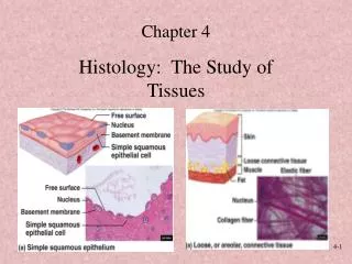
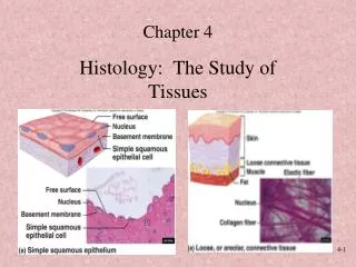
General Properties of Connective Tissue 1. One of the four basic types of tissues (epithelium, connective tissue, muscle, and nervous tissue) 2. Composition: • cells (fibroblasts and others), • fibers and ground substance (extracellular matrix) 3. Functions: • Architectural framework of the body • Bind together and provide mechanical support for othertissue (metabolic, defense, transport, storage) • Wound repair / inflammatory response
Connective Tissue • Extracellular Matrix Fibers – collagen & elastic “Ground substance” • Cells Fixed: Fibroblasts Adipocytes “Tissue macrophages” Free: Immune cells (lymphocytes) Inflammatory cells (neutrophils & activated macrophages)
Fibers in Connective Tissue • Collagen • most abundant protein in human body (up to 30% dry weight) • multiple types:fibril-formingorfibril-associated(in skin, tendon, cartilage, bone, dentin, blood vessels);cross-linked networks(in all basement membranes) • Reticular Fibers– specialized type of collagen(Type III; reticulin)associated with smooth muscle in organs subjected to changes in volume, forms the stroma in lymphatic and hematopoietic organs • Elastic Fibers–thin fibers or fenestrated sheets composed of various glycoproteins, including the protein elastin, providing elastic properties to tissues that experience repeated deformation (in skin, blood vessels, lung, bladder)
Major Collagen Fiber Types (out of at least 20) TYPE LOCATION FUNCTION Fibril-forming collagens (these are visible) I (most abundant) Skin, tendon, bone, dentin Resistance to tension II Cartilage, vitreous of eye Resistance to pressure III (reticulin) Skin, blood vessels, organs Structural framework and stability Network-forming collagens IV All basement membranes Support and filtration Fibril-associated collagens with interrupted triple helices (FACIT) multiple types Assoc. w/ type I and II fibrils Fibril-fibril / fibril-ECM binding Anchoring filament collagens VII Epithelia Epidermis to basal lamina
Collagen fibers viewed by light microscopy Trichrome H&E
Collagen Fibers vs. Fibrils H & E fibrils fibers
Collagen Synthesis DISULFIDE BONDS ALDOL CONDENSATION (aka tropocollagen)
Site of Lys/OH-Lys aldol condensation (Lys only at ends of molecules) • Heavy metal stains preferentially fill gap regions 67nm banding caused by overlaps at ends of molecules
FACIT: Fibril-Associated Collagens with Interrupted Triple helices • Triple helices interrupted by non-helical domains • Retain propeptides at ends • Do not aggregate into large fibrils • Bind collagen fibrils to each other and/or the ECM Type IX Collagen (left) I • Binds type II Fibrils to the ECM • Globular N-terminus interacts with ECM • Heparin-SO4 at kink interacts with ECM • Helical region interacts with type II fibril Type VI Collagen (right) Bundles type I fibrils into FIBERS Binds fibrils via helical domains
Reticular (Reticulin) Fibers • Form a delicate supporting frameworkfor highly cellular tissues (endocrine glands, lymph nodes, liver, bone marrow, spleen, smooth muscle). • Composed mainly of Type III collagen, with a carbohydrate moiety that reduces Ag+ to metallic sliver = argyrophilic. • Special stain: silver impregnation to visualize. • Thinner than type I collagen(Type III fibrils are 30-40 nm diameter; type I fibrils are ~200 nm diameter)
Reticular Fibers (type III collagen) • made by reticular cells (specialized fibroblasts) and vascular smooth muscle cells
Clinical disorders resulting from defects in collagen synthesis
Ehlers-Danlos Syndromes • A series of genetic diseases with faulty assembly of collagens. • Hyperextensible skin and hypermobile joints • In some forms (e.g., type IV), weakness in blood vessels or intestines are life threatening.
Noncollagen Components of the Extracellular Matrix • Elastin • “Ground substance” • Glycosaminoglycans (GAG’s) • Proteoglycans • Multiadhesive matrix proteins • laminin • fibronectin
Elastic Fibers LM:Visualized by selectively staining withWeigert’s, resorcin-fuchsin, or aldehyde-fuchsin EM:Consist of amorphous core ofelastin surrounded by microfibrillar glycoprotein,fibrillin(8-10nm). Elastin:is rich in glycine and proline, but it containslittle or no hydroxyproline and hydroxylysine .uniquely containsdesmosine and isodesmosine,which are thought to cross-link the molecules into anetwork of randomly coiled chains. This cross-linkingis responsible for its rubber-like properties. Confers elasticity:present in large amounts in ligaments, lung,skin, bladder, and walls of blood vessels. Marfan Syndrome:defect in elastic fiber synthesis; reduced elasticity in skin and lungs, skeletal defects (bones are longer and thinner than usual), cardiovascular complications (aneurism, valve prolapse)
Network of elastin molecules can stretch and recoil like a rubber band
Elastin appears amorphous (not fibrillar) in the electron microscope E=elastin C, collagen fibrils M/L=microfibrils of fibrillin, a scaffolding glycoprotein involved elastin deposition Marfan Syndrome: defect in fibrillin gene, results in weakened elastic fibers
Elastic and Collagen Fibers elastin stain (“Weigert’s”, “aldehyde fuchsin”, “Verhoeff”): elastic fibers are purple/black collagen fibers stain orange/pink or blue/green depending on other stains used (von Gieson’s or trichrome, respectively) H&E stain: collagen stains orange/pink; elastic fibers stain glassy red (generally only visible if in HIGH abundance)
Ground Substance of the Extracellular Matrix (ECM) • Glycosaminoglycans (GAG) • linear (unbranched) polysaccharides, e.g. heparan sulfate, condroitin sulfate, keratan sulfate, hyaluronic acid • very hydrophilic due to abundant negative charges (e.g. SO4- groups). • except for hyaluronic acid, are usually bound covalently to protein core as part of a proteoglycan • Proteoglycans • core protein + GAG side chains (like a bottle brush) • bind cells, other proteins, and/or ECM components • Multiadhesive glycoproteins • small glycosylated proteins containing NUMEROUS binding sites to cells, signaling molecules, and other ECM components • e.g. fibronectin and laminin: important for adhesion of epithelial cells to the basal lamina via transmembrane integrin receptors.
Basement Membrane – Collagen Types IV, VII, and III • Basement membranes are sheets of extracellular matrix proteinslocated at the interface of parenchyma (epithelia, endothelia, muscle, nerves, adipocytes) and connective tissue / ECM. • Main constituentsare glycosaminoglycans(heparan sulfate),fibrous proteins(collagen typesIV,VII, III), structural glycoproteins fibronectin, laminin and entactin. • This is NOT a plasmamembrane.
Basement membranes vary in thickness Thick Thin -- requires special stain to visualize BM BM BM Intestinal glands, PAS trachea, H&E PAS reacts with carbohydrate-rich molecules such as perlecan, laminin and type III collagen associated with the basement membrane.
A closer look at the basement membrane: What appears as ONE layer by LM is actually THREE layers when viewed by EM hemidesmosomes • lamina lucida (LL) or rara 10-50 nm • lamina densa (LD) 20-300 nm (type IV collagen) basal lamina fibroreticular lamina • Fibroreticular lamina (FL) merges with underlying CT (type III* and type VII collagen fibrils) LL LD FL Connective tissue *so, basement membranes can also be visualized with silver stain So, the “basement membrane” is the basal lamina + the fibroreticular lamina
Tying it all togetherInteractions of many proteins tether cell to the underlying connective tissue: • Cell to basal lamina… • Hemidesmosome • Type IV collagen • Integrin/laminin • Basal lamina to underlying connective tissue: • Type IV collagen • Type VII collagen • Fibrillin • Type III collagen lamina rara lamina densa anchoring fibril (collagen VII) reticular fibril (collagen III)
Cells in Connective Tissue • Fibroblasts • Adipose (fat) cells • Tissue Macrophages** • Mast cells** • Lymphocytes & Plasma Cells (differentiated B-cells)** • “Leukocytes”** Fixed(permanent residents) Free(transient residents) (specifically, neutrophils, eosinophils, & basophils) ** derived from hematopoietic stem cells and involved in immune function and inflammation
Fibroblastsare the most common cells in connective tissue • Synthesize and secrete components of the ECM: fibers and ground substance. • Active and quiescent stages(when quiescent sometimes called fibrocytes or mature fibroblasts). • Synthesize growth factors. • Rarely undergo cell divisionunless tissue is injured, which activates the quiescent cells. • Play a major role in the process of wound healingand respond to an injury by proliferating and enhanced fiber formation.
Single, large lipid droplet Adipocytespredominate in adipose tissue • Very active cells with many functions: • Triglyceride storage and glucose metabolism (insulin and glucagon receptors) • Secretion of many bioactive molecules: • leptin (regulates satiety) angiotensinogen (blood pressure) steroids (glucocorticoids & sex hormones) • growth factors (e.g. insulin-like growth factor, tumor necrosis factor ) cytokines (e.g. interleukin-6) White (common, yellow, unilocular) adipose tissue stained with Masson’s trichrome
Adipocytes Lipid (fat) droplet Nucleus Capillaries
Present in newborns (and hibernating mammals) and involved in thermoregulation Brown (Multilocular) Adipose Tissue Mitochondria of brown fat cells express uncoupling protein (UCP), which “short circuits” the electron transport chain producing HEAT rather than ATP. white brown
Cells of the blood • Erythrocytes(red blood cells, RBC) • Leukocytes (white blood cells, WBC) • Granulocytes (with specific granules) • Neutrophil (~60% of WBC) • Eosinophil (~5% of WBC) • Basophil (<1% of WBC) • Agranulocytes (without specific granules) • Lymphocyte (B-cell, T-cell) (~25% of WBC) • Monocyte (~10% of WBC) (also in the blood are platelets, which are small, membrane-bound cell fragments involved in blood clotting… to be discussed later)
Human blood smear, with RBCs, WBCs and platelets Platelets RBC Lymphocyte Neutrophil
Erythrocyte (red blood cell, RBC) • Size and shape: • biconcave disk, 8 µm diameter, 2m at thickest point, 1 m at thinnest • flexible: RBC’s normally bend to pass through small capillaries • LM appearance in smear: Pink circle with light center (center is thinner because of the biconcave shape). No nucleus. • Function: • Transport of oxygen and carbon dioxide • bound to hemoglobin (oxyhemoglobin and carboxyhemoglobin) • majority of CO2 transported as HCO3- • pH homeostasis • carbonic anhydrase: CO2 + H2O HCO3- + H+ • HCO3- / Cl- antiporter : exchanges HCO3- for extracellular Cl-
Red blood cells in a blood smear RBC Platelet
Platelets (thrombocytes) • Shape, size, and origin: Small, biconvex disks, 2-3 µm in diameter. Non-nucleated cell fragments derived from cytoplasm of a very large cell, the megakaryocyte, in bone marrow. Platelets have a life span of about 10 days. • LM appearance in smears: Small basophilic fragments, often appearing in clusters. • Function: Platelets initiate blood clots.
RBC & platelet, TEM RBC Platelet
Neutrophil • Specific granules • Type IV collagenase (aids migration) • Lactoferrin (sequesters iron) • Phospholipase A2 (leukotriene synthesis) • Lysozyme (digests bacterial cell wall) • Non-specific granules (lysosomes) • Lysozyme • Acid hydrolase • Myeloperoxidase • Elastase • Granulocyte with specific and non-specific granules, ~60% of WBCs • LM appearance in smear: About 9-12 µm in diameter (thus larger than RBC). Nucleus long and multi-lobed (usually 2-4 lobes). 3. Cytoplasm has small, neutrally stained specific granules. Non-specific granules are azurophilic. 4. Function: Primarily antibacterial • Neutrophils leave the blood and follow chemotaxic signals to sites of wounding or other inflammation, and phagocytose foreign agents such as bacteria. Pus is composed largely of dead neutrophils.
Two neutrophils in a blood smear LM appearance in smear: About 9-12 µm in diameter (thus larger than RBC). Nucleus long and multi-lobed (usually 2-4 lobes). Cytoplasm has small, neutrally stained specific granules. Non-specific granules are azurophilic.
Neutrophil, transmission electron micrograph TEM appearance: Multi-lobed nucleus and numerous specific granules and lysosomes (=azurophilic granules in LM). Specific granule Lysosome (=azurophilic granule)
Neutrophils in tissue: • Enter connective tissue from blood vessels as the “first wave” in acute inflammatory responses • Small cells with multi-lobed, heterochromatic nuclei (aka “polymorphonuclear neutrophils”, “PMNs”, “polys”) • Primary function: anti-bacterial (are phagocytic like mphages, but SHORT-lived and NOT antigen presenting)
Eosinophil • Specific granules • Major basic protein • Eosinophilic cationic protein • Neurotoxin • Histaminase • Non-specific granules (lysosomes) • Lysozyme • Acid hydrolase • Myeloperoxidase • Elastase • Granulocyte with specific and non-specific granules, 3-5% of WBCs • LM appearance in smear: About 10-14 µm in diameter. Bilobed nucleus. The cytoplasm has prominent pink/red specific granules (stained with eosin dye). If the smear is not stained properly, the granules may be brownish. • Function: • Anti-parasitic activity • Mediators of inflammatory/allergic responses in tissues • Inactivate leukotrienes and histamine secreted by basophils • Engulf and sequester antigen-antibody complexes • Inflammatory stimulus increases production/release of eosinophils from bone marrow, whereas inflammatory suppression decreases eosinophil numbers in peripheral blood. • But, they also secrete PRO-inflammatory chemokines AND they can degranulate inappropriately to cause tissue damage (as in reactive airway disease)
Eosinophil in a human blood smear LM appearance in smear: About 10-14 µm in diameter. Bilobed nucleus. The cytoplasm has prominent pink/red specific granules (stained with eosin dye). If the smear is not stained properly, the granules may be brownish.
Eosinophil, transmission electron microscopy externum internum TEM appearance: The specific granules are ovoid in shape, and contain a dark crystalloid body composed of major basic protein (MBP), effective against parasites. The rest of the granule contains other anti-parasitic substances and histaminase. The cytoplasm also contains lysosomes (=azurophilic granules).
Eosinophils can also migrate into connective tissue (often seen in chronic allergies or inflammatory diseases)