SCANNING ELECTRON MICROSCOPY(SEM) METHODS IN STUDY OF MICRO OBJECTS
SCANNING ELECTRON MICROSCOPY(SEM) METHODS IN STUDY OF MICRO OBJECTS. GAQA S.G. MANAKA M.C. MOIPOLAI T.B. THABETHE B.S. SUPERVISOR: O.ORELOVITCH FLEROV LABORATORY OF NUCLEAR REACTIONS, CENTER OF APPLIED PHYSYSCS. LAYOUT. AIMS AND OBJECTIVES INTRODUCTION GENERAL VIEW OF SEM JSM-840

SCANNING ELECTRON MICROSCOPY(SEM) METHODS IN STUDY OF MICRO OBJECTS
E N D
Presentation Transcript
SCANNING ELECTRON MICROSCOPY(SEM) METHODS IN STUDY OF MICRO OBJECTS GAQA S.G. MANAKA M.C. MOIPOLAI T.B. THABETHE B.S. SUPERVISOR: O.ORELOVITCH FLEROV LABORATORY OF NUCLEAR REACTIONS, CENTER OF APPLIED PHYSYSCS
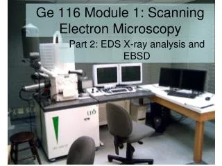
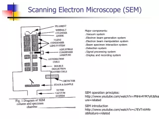
LAYOUT • AIMS AND OBJECTIVES • INTRODUCTION • GENERAL VIEW OF SEM JSM-840 • HOW SEM WORKS • GENERAL VIEW OF ION SPUTTER • EXPERIMENTAL PROCEDURE • RESULTS AND DISCUSSION • CONCLUSION
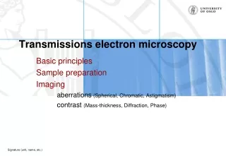
AIMS AND OBJECTIVES • Learning the principles of SEM • Study the methods of sample investigations with the use of SEM
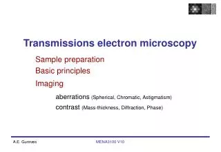
INTRODUCTION • What is SEM(Scanning electron microscopy) SEM is a type of electron microscope that images the sample by scanning it with high energy beam of electrons . The signals obtained from electron-sample interaction reveal information about the sample including external morphology (texture), chemical composition, and crystalline structure and orientation of materials making up the sample. • Why SEM . SEM is indispensable when observation of objects with the size less than the wavelength of light is required
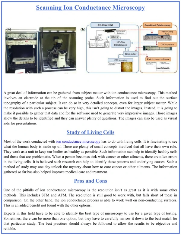
HOW DOES SEM WORK • TV signal from SEM is transferred to analog-digital converter (ADC) • It is then processed by special system and shown on the computer monitor as digital picture in PCX format.
ION SPUTTER The specimen stage with the membrane samples are placed on the specimen stand under the bell jar The membrane samples are covered with gold by setting the air pressure under the bell jar to 0.2 torr, voltage to 1.2kV and the current to 10 mA for two minutes
EXPERIMENTAL PROCEDURE • The specimen stage was cleaned with acetone. • The membrane samples were mounted on the specimen stage using silver conductive glue. • The specimen stage together with the mounted membrane samples were placed on the ion sputter to cover with gold. • The membrane samples were placed in the scanning electron microscope for characterization.
RESULTS AND DISCUSSION • Without covering there is a surface charge • It is due to the surface resistance of the polymer membrane to the secondary electrons. • The surface of the sample is not thermo conductive.
CONTINUED • With covering there is less surface charge • the surface of sample is electro conductive to minimize charging from the incident beam • the surface of sample is thermo conductive to minimize local heating • the material of sample has a high atomic number to increase the coefficient of secondary electrons .
CONTINUED • With tilting a better view of the topography is achieved
CONTINUED • A more better view of the surface and pores is achieved at increased tilting angles.
METAL MEMBRANE • Metal membrane • Conductive • Surface topography can still be achieved without covering with gold.
SODIUM CHLORIDE • Covered with gold by rotating the sample twice at an angle of 90 degrees
CONCLUSION • The principles of SEM and methods of sample preparation were learned • Membrane samples covered with gold showed no surface charge and the uncovered membrane samples showed a surface charge • A better view of the topography was achieved when tilting the sample.
ACKNOWLEDGEMENTS • NRF and DST for funding • Organisers of the student summer practice • JINR(University Centre) for the study opportunity • Project supervisor, Dr. Oleg Orelovitch for his unending support through out the project
REREFENCE • D. Pease, Histological Techniques for Electron Microscopy, Academic Press, New York and London, 1960 • J. Goldstein, H. Yakowetz, PRACTICAL SCANNING ELECTRON MICROSCOPY, Plenum Press, New York and London, 1975
THANK YOU • QUESTIONS