Download
1 / 81
810 likes | 1.07k Vues
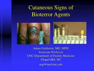
Cutaneous Signs of Bioterror Agents. Adam Goldstein, MD, MPH Associate Professor UNC Department of Family Medicine Chapel Hill, NC aog@med.unc.edu. Objectives. Improve ability to: diagnose and manage cutaneous illness associated with suspected cases of bioterror

E N D
Cutaneous Signs of Bioterror Agents Adam Goldstein, MD, MPH Associate Professor UNC Department of Family Medicine Chapel Hill, NC aog@med.unc.edu
Objectives • Improve ability to: • diagnose and manage cutaneous illness associated with suspected cases of bioterror • Anthrax, plague, tularemia, smallpox, mustard gas
Why worry? • “Subnational attacks using genetically engineered organisms are inevitable” • “Biologic agents now join nuclear agents” • Deaths • 1 KT H-BOMB .6M – 2M • 100 Kg ATX 1M – 3M (Stansfield Turner, CIA, 2001)
Anthrax • Anthrakos = ‘coal’ b/c of black eschar • B. anthracis is gram-positive sporulating bacillus • Spores are resistant to heat, cold, drying, & chemical disinfection • Anthrax is endemic in western Asia (Iran Turkey Afghanistan,) & western Africa (McGovern, Elect Text Dermatol, 1999)
Anthrax • Spores viable for yearstop 6 cm of soil & in animal products • Disease transmitted from infected animals or products via skin abrasions > 90% of cases • Goats > sheep > cattle > horses > pigs > dogs
Anthrax • Burn dead animals, not buried, to prevent long-term environmental contamination
History of Anthrax • 1500 B.C. -- Fifth/sixth Egyptian plagues, ? Anthrax • 1600s -- "Black Bane," ? anthrax, kills 60,000 cattle • 1876 -- Koch confirms bacterial origin of anthrax • 1880 -- Immunization of livestock against anthrax • 1915 -- German agents in U.S. inject horses/cattle with anthrax on way to Europe during WW I • 1937 -- Japan starts biological warfare program • 1942 -- Britain experiments with anthrax • 1943 -- U.S. begins developing anthrax weapons • 1945 -- Anthrax outbreak in Iran kills 1 million sheep
Historical • 1950s and '60s -- U.S. biological program continues • 1969 -- Nixon ends U.S. offensive biological program. • 1970 -- Anthrax vaccine approved by U.S. FDA • 1972 -- International convention outlaws development or stockpiling of biological weapons • 1978-80 -- Human anthrax epidemic strikes Zimbabwe, infecting > 6,000 and killing 100 • 1979 -- Aerosolized anthrax spores released at Soviet military facility, killing 68 • 1991 -- U.S. troops vaccinated for Gulf War I • 1990-93 -- Terrorists release anthrax in Tokyo; no injuries
Historical • 1995 -- Iraq produced concentrated anthrax in biological weapons program • 1998 -- U.S. approves anthrax vaccinations for all military • 2001 -- Letter with anthrax spores mailed to NBC one week after 9/11 terrorist attacks on Pentagon & WTC. Several die after inhaling.
Anthrax pilot plant used to produce billions of anthrax spores at Fort Detrick, Md. U.S. ended offensive biological weapons research in 1969
Al Hakam, Iraq's major facility for production of biological agents. Plant destroyed by Iraqi workers in 1996.
Pulmonary Anthrax • Wool-sorter’s disease • 18 cases reported in U.S. 1900-1980 • Symptoms: vague prodrome with fever, malaise, myalgias and cough • Within days- rapidly developing precordial discomfort, cyanosis, stridor, diaphoresis, moist rales, pleural effusion and death
X-ray findings: hemorrhagic mediastinitis, but not true pneumonia; widened mediastinum
Cutaneous Anthrax Incubation period 7 days (1-12 range) • Initial painlesspapule (head, neck, extremity) • May resemble spider bite and may itch • Surrounding erythema & edema • Vesicle or bulla rapidlyevolves • Painless hemorrhage & necrosis • Fluid becomes black • Lesion ulcerates & develops black eschar with surrounding edema • Pearl-like satellite vesicles may occur
Lesions progress from: papule - erythema - vesicle - necrosis - ulcer - eschar with or without antibiotic therapy progression d/t toxin Lesions may be solitary or multiple (same part of body) Occasionally associated: Tender lymphadenopathy Fatigue Fever and/or chills (Caruscci, JAAD 2001) Cutaneous Anthrax
Cutaneous Anthrax - Painless Lesions • Surrounding edema or regional lymphadenopathy may be painful. • Debridement of skin lesions notindicated b/c risk of spreading infection
Cutaneous Anthrax: Diagnosis • Notify local Health Department • Before doing diagnostic tests • Mask not required & personnel not at risk • Disease acquired through contact with spores, not active bacteria
Diagnosis • Swab exudates for Gram stain & culture (fresh vesicles) • 4-mm punch biopsy full-thickness (through entire dermis) • permanent sections • immunohistochemistry studies • polymerase chain reaction (PCR) • A second punch biopsy for Gram stain, bacterial, fungal & atypical mycobacterial cultures • Send clinical history (& lesion picture if possible) • Negative bx does not r/o cut. anthrax b/c skin lesions caused by toxins
Diagnosis • Draw 5 mL of blood in red-topped tube • Transfer to laboratory for isolation of serum & subsequent storage at –70°C- label tube: “Anthrax serology. • Store serum at –70°C for special pick-up.” • Draw 5 mL of blood into a purple-topped tube • Refrigerate • Hold for pick-up- PCR diagnostic tests by CDC
Pruritic and papular arthropod bites Brown recluse and other spider bites Pustular diseases Antiphospholipid antibody syndrome ulcers Aspergillosis Coumadin or heparin necrosis Ecthyma gangrenosum Cutaneous leishmaniasis Mucormycosis Plague Rickettsial pox Staphylococcal & streptococcal ecthyma Tropical ulcer Tularemia Typhus, scrub and tick Differential Diagnosis:(eschar/ulceration)
Chancroid Glanders Herpes simplex Cutaneous leishmaniasis Lymphogranuloma venereum Melioidosis Cutaneous nocardiosis Plague Sporotrichosis & other deep fungal diseases Staphylococcal & streptococcal adenitis Tuberculosis Tularemia Differential Diagnosis: (ulceroglandular)
Treatments http://www.bt.cdc.gov/agent/anthrax/index.asp
Treatments • If suspected anthrax, begin appropriate tx • Tx regimen differs by symptomatology (systemic or localized), location (extremity vs head/neck), edema (extensive or not) • If systemic signs, head or neck location, or extensive edema, IV therapy indicated
Treatment for cutaneous anthrax patients without systemic symptoms, not located on the head or neck, not with extensive edema, & not in children younger than 2 years Category Initial oral therapy Duration (days) Adults Ciprofloxacin, 500 mg bid 60 or doxycycline, 100 mg bid Children Ciprofloxacin, 15 mg/kg q12h 60 (not to exceed 1 g/d) or doxycycline: >8 y o, >45 kg, 100 mg q12h; all other children, 2.2 mg/kg q12h Pregnant Ciprofloxacin, 500 mg bid (preferred) 60 or doxycycline, 100 mg bid Immunocomp Same 60
Treatment of cutaneous anthrax with systemic symptoms, extensive edema, involving the head or neck, or children < than 2 yo (same as for inhalational anthrax) Category IV therapy Duration (days) Adults Ciprofloxacin, 400 mg q12h, IV initially, oral or doxycycline,100 mg q12h, when stable, 60 days and 1-2 additional agents Children Ciprofloxacin, 10 mg/kg q12h IV initially, oral (not to exceed 1 g/d)| or when stable, 60 days doxycycline: >8 y old and >45 kg, 100 mg q12h; all other, 2.2 mg/kg q12h and 1-2 additional agents Pregnant & Same as for nonpregnant Same Immunocom and immunocompetent adults & children
Usually painful Bites from spiders of the genus Loxoceles begin as pale ecchymotic lesions that rapidly turn purple. Lesions may ulcerate and develop necrotic centers Borders are irregular, ill-defined and without the significant surrounding edema. Spider bites: Usually painful