Download
1 / 111
1.11k likes | 1.13k Vues
BASIC STRUCTURE & FUNCTION OF THE SKIN AND CUTANEOUS SIGNS, PRINCIPLES OF DERMATOLOGIC DIAGNOSIS. Oktay Taşkapan, MD Professor of Dermatology & Allergy, Chair, Department of Dermatovenereology Yeditepe University, Faculty of Medicine İstanbul.

E N D
BASIC STRUCTURE & FUNCTION OF THE SKIN AND CUTANEOUS SIGNS, PRINCIPLES OF DERMATOLOGIC DIAGNOSIS Oktay Taşkapan, MD Professor of Dermatology & Allergy, Chair, Department of Dermatovenereology Yeditepe University, Faculty of Medicine İstanbul
To Sam Shuster, who showed me that there is more to dermatology than meets the eye. J.L.Burton,MD
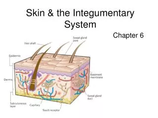
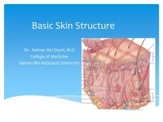
Epidermis • Dermis • Subcutis (SC tissue) • Epidermal appendages (adnexa)
The skin is the largest organ in the body • It separates thebody's internal environment from the external environment • A channel of communication with the outside world; protects the body from water loss; usesspecialized pigment cells, called melanocytes, to protect the body from ultraviolet radiation;participates in calcium homeostasis by contributing to the body's supply of vitamin D; and helpsregulate body temperature and metabolism.
EMBRYOLOGY • First weeks of life Periderm • Third month Adnexal structures • Epidermal stem cells Epidermis, sweat gland epithelium and hair epithelium • Epidermis and epidermal appendages (hairs, sebaceous glands, eccrine glands, apocrine glands and nails): Ectoderm • Dermis, SC fat, vessels, muscles: Mesoderm • Nerves and melanocytes: Neuroectoderm and neural crest
The skin is composed of diverse cell types of both ectodermal (e.g. keratinocytes, melanocytes, Merkel cells, neurons) and mesodermal (e.g. fibroblasts, hematopoietic cells such as Langerhans cells, endothelial cells) lineages.
The epidermis is formed mainly by keratinocytes • They synthesize keratin, a filamentous protein that serves a protective function • Palms and soles: Epidermis 1.5 mm • Eyelid: Epidermis 0.1 mm
Keratinocytes • Keratin, a complex filamentous protein, is the surface coat of the epidermis, and the structural component of hair and nails • Keratins are critical for normal epidermal function • Basal layer (stratum germinativum), malpighian or prickle layer (s.spinosum), granular layer (s.granulosum: Keratohyaline granules), s.lucidum and horny layer (s.corneum) • Tonofilaments and desmosomes
Programmed process of maturation resulting in death Terminal differentiation • The epidermal transit time is about one month • Keratinocytes play an active role in the immune function • They secrete cytokines and inflammatory mediators • They express ICAM-1 and MHC class II molecules on their surface
Keratinocytes • Melanocytes • Langerhans cells • Merkel cells
Melanocytes • Pigment producing (dendritic) cells of the epidermis / neural crest origin • One melanocyte a great number of keratinocytes (“epidermal melanin unit”) • Basal layer at a frequency of approximately 1 in every 10 basal keratinocytes (sun protected trunk epidermis) • Racial differences in skin colour are caused by the number, size and distribution of the melanosomes (pigment granules within keratinocytes) • Within keratinocytes, melanin forms a photoprotective cap over the nucleus
Melanin is formed in melanozomes • Melanosomes are travelled along dendrites • Melanosomes are injected into the cytoplasm of the neighbouring keratinocytes
Langerhans cells • Found scattered among keratinocytes of the S.spinosum • 3-5 % of the cells in this layer • Connected to adjacent keratinocytes by the desmosomes • Birbeck granules • Bone marrow origin • Recognition, uptake, processing and presentation of antigens to sensitized T lymphocytes [“Antigen Presenting Cells (APC) in cutaneous immunity]
Merkel Cells • Located above the basement membrane zone • Direct connections with adjacent keratinocytes by desmosomes • Synaptic contacts with somatosensory afferents • Sense of light touch discrimination of shapes and textures (act as receptor for mechanical stimuli).
Dermis: Cells, fibers and ground substance • The constituents of the dermis are mesodermal in origin except for nerves which derive from the neural crest • 0.3 mm on the eyelid, 3.0 mm on the back • The principal component of the dermis is collagen (fibrous proteins) • The fibroblast synthesizes the procollagen molecule, reticulum fibers, elastic fibers and the ground substance (extracellular matrix): Connective tissue • Ground substance: Sulfated acid mucopolysaccharide (chondroitin sulfate, dermatan sulfate, electrolytes) • Histiocytes (macrophages), fibroblasts, lymphocytes, plasma cells, mast cells
Collagen is the major stress-resistant material of the skin • Elastic fibers have a role in maintaining elasticity • Papillary dermis: The thin upper layer, composed of thin, haphazardly arranged collagen fibers • Reticular dermis: The thicker lower layer, extends from the base of the papillary layer to the SC tissue and is composed of thick collagen fibers • Dermal papillae – Rete ridge
Dermoepidermal junction • DEJ is formed by the “basement membrane zone” • A structural support holding the epidermis and dermis together • It is considered a porous semipermeable filter which permits exchange of cells and fluid
Epidermal appendages: Adnexa • Hair follicles, sebaceous glands (pilosebaceous units), eccrine and apocrine glands, nails
Hair follicles • Infundibular segment (from its surface opening to the entrance of the sebaceous duct) • Isthmus (between the sebaceous duct and the insertion of the arrector pili muscle) • Hair bulb
On the scalp, anagen, active growth phase, lasts about 3-5 years • 85-90 % of all scalp hairs are in the anagen phase (0.37 mm / day) • Catagen (involution phase): 2 weeks • Telogen (resting phase): 3-5 months • Each follicle functions as an independent unit
Sebaceous glands • Greatest abundance on the face and scalp • Distributed throughout all skin sites except the palms and soles • Mostly associated with hair follicles (pilosebaceous unit) • The main function of the sebaceous glands is to provide lipids, which lubricate the hair shaft and, along with lipids produced by the epidermal cells, maintain a lipid film on the skin surface
Eccrine sweat units • Most abundant on the palms, soles, forehead, and axillae • Cholinergic innervation • Heat and emotional stress • Thermoregulation, acid mantle
Apocrine units • Open into the infundibular portion of the hair follicle • Adrenergic innervation • Apocrine sweat is odorless until it reaches the skin surface, where it is altered by bacteria • Generally confined to axillae, areolae, anogenital region, external auditory canal, and eyelids • Its function is unknown
Nails • Act to assist in grapping small objects and in protecting the fingertip • Matrix keratinization leads to the formation of the nail plate • Fingernails: 0.1 mm/day
Dermal vasculature • The subpapillary plexus (upper horizontal network) • Lower horizontal (deep) plexus is found at the dermal-SC interface and is composed of larger blood vessels • Associated with the vascular plexus are dermal lymphatics and nerves
Muscles • Smooth muscle (SM): Arrectores pilorum, tunic dartos of the scrotum, in the areolas around the nipples • Anogenital skin: Scattered SM throughout the dermis • The muscularis of dermal and SC blood vessels • Glomus bodies: Specialized aggregates of SM cells found between arterioles and venules (prominent on the digits and at the lateral margins of the palms and soles) • Superficial muscular aponeurotic system (SMAS): Striated muscle of neck and face
Nerves • The dermis is rich in nerves (neurovascular bundle) • Touch and pressure are mediated by Meissner corpuscles found in the dermal papillae (particularly on the digits, palms and soles) and Vater-Pacini corpuscles located in the deeper portion of the dermis of weight bearing surfaces and genitalia • Temperature, pain and itch sensation are transmitted by unmyelinated nerve fibers • Postganglionic adrenergic fibers of ANS: Vasoconstriction, apocrine gland secretions, and contraction of arrector pili muscles • Cholinergic fibers: Eccrine sweat secretion