Positron Emission Tomography
Positron Emission Tomography. P E T. Positron Emission Tomography. Introduction. What is PET? Revision of beta decay and isotopes. How a positron annihilates with an electron. How PET works. Medical uses for PET scanning. brain. heart. kidney. bladder. Introduction.

Positron Emission Tomography
E N D
Presentation Transcript
Positron Emission Tomography www.howpetworks.com
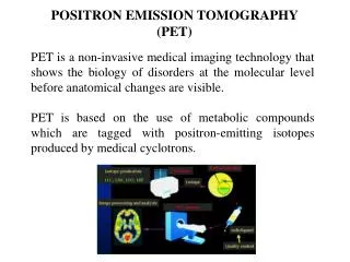
P E T Positron Emission Tomography Positron Emission Tomography
Introduction • What is PET? • Revision of beta decay and isotopes. • How a positron annihilates with an electron. • How PET works. • Medical uses for PET scanning. Positron Emission Tomography
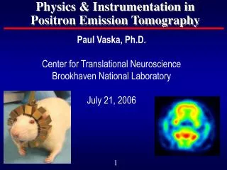
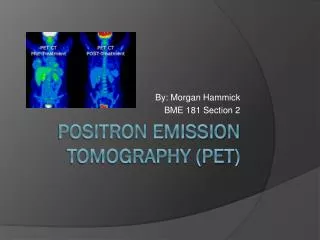
brain heart kidney bladder Introduction What organs appear dark in this scan? Positron Emission Tomography
What Is PET? Positron Emission Tomography
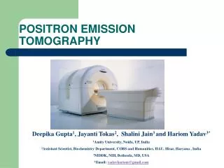
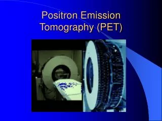
Having a PET Scan • Minute amount of radioactive isotope injected into patient. • Patient lies still 30 to 60 minutes in a chamber like the one illustrated. • Scan is looked at by an imaging specialist • Results passed to patient’s doctor. Positron Emission Tomography
Revision: Beta Decay • When a nucleus is unstable, there are three ways it can decay. It can emit: • An alpha particle • A beta particle • A gamma ray or Positron Emission Tomography
Beta-minus decay (β -) Revision: Beta Decay Beta-plus decay (β +) Positron Emission Tomography
Revision: Isotopes Atoms of the same atomic number but with different mass numbers (i.e. with different numbers of neutrons) are called isotopes. Example: carbon atoms always have 6 protons in the nucleus, but there are different isotopes, each of which has a different mass number. These are called carbon-11, carbon-12 etc. Positron Emission Tomography
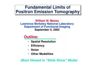
Revision: Isotopes • Isotopes can be plotted on a graph like this. • Isotopes that lie on this line are stable. • Isotopes that are above or below the line are unstable or radioactive. Positron Emission Tomography
How PET works: Positron-electron Annihilation When a positron and electron collide, they annihilate each other and emit gamma rays at 180 degrees to each other. Positron Emission Tomography
How PET Works: Cyclotron Positron Emission Tomography
How PET Works: Labelling and Tracers • Positron-emitting isotopes label molecules. • An image can be acquired showing the location of these molecules. • The most common molecule labelled is Fluoro-deoxyglucose (FDG), which behaves almost exactly like glucose in the body. Positron Emission Tomography
Applications of PET:Cancer Diagnosis PET scanning can be used to find out where cancerous tumours are, and how far they have spread in the body. This is very important in choosing the best treatment for the patient. tumour Positron Emission Tomography
Applications of PET:Cancer Diagnosis • 18-FDG behaves almost exactly like glucose. • Cancer cells use up a lot of glucose as they rapidly divide. • Can be used to stage the disease, and decide the best treatment. Positron Emission Tomography
Applications of PET:Alzheimer’s Disease • The most common type of dementia • Can gradually destroy people's memory. • Patients with Alzheimer’s have protein deposits in the brain called Amyloid plaques. • PET can be used to detect the presence of Amyloid plaques. Positron Emission Tomography
Applications of PET:Alzheimer’s Disease Normal Alzheimer’s PET Scanning with 18-FDG (made with Fluorine-18) Positron Emission Tomography
Applications of PET:Alzheimer’s Disease Normal Alzheimer’s PET Scanning with 11C-PIB (made with Carbon-11) Positron Emission Tomography
Applications of PET:Alzheimer’s Disease Normal Alzheimer’s PET Scanning with 11C-PIB (made with Carbon-11) Positron Emission Tomography
Applications of PET:Heart Disease PET can be used to work out whether or not it is worth performing an operation on heart muscle. Positron Emission Tomography
Applications of PET:Heart Disease After a heart attack, heart muscle can be: • Stunned • Hibernating • Dead Heart muscle that is alive is often called viable. Positron Emission Tomography
Summary • PET scanning is one of the most important medical applications of radioactivity • It involves a type of beta decay called β+ or positron • A positron emitting chemical is injected into the patient, who is then imaged • Positron’s don’t travel far inside a patient, but annihilate when they meet an electron • The resulting gamma rays are detected by a PET scanner. Positron Emission Tomography