Positron emission tomography in oncology
300 likes | 609 Vues
Positron emission tomography in oncology. Prof. Dr. Alex Maes AZ Groeninge Kortrijk KUL Campus Kortrijk. RIZIV-terugbetaalde indicaties van PET-onderzoeken. Geïsoleerde longnodule van onbekende aard Vermoeden recidief of residuele massa ter hoogte van hersenen, mond of farynx

Positron emission tomography in oncology
E N D
Presentation Transcript
Positron emission tomography in oncology Prof. Dr. Alex Maes AZ Groeninge Kortrijk KUL Campus Kortrijk
RIZIV-terugbetaalde indicaties van PET-onderzoeken • Geïsoleerde longnodule van onbekende aard • Vermoeden recidief of residuele massa ter hoogte van hersenen, mond of farynx • Opstellen staging bij • Longgezwel (niet kleincellig) • Slokdarmgezwel • Pancreasgezwel • Maligne melanoom vanaf stadium IIc (AJCC-classificatie) • Lymfoom van intermediaire of hoge grading
RIZIV-terugbetaalde indicaties van PET-onderzoeken • Vermoeden recidief of residuele massa bij • Maligne melanoom • Longtumor (niet kleincellig) • Colorectale tumor • Pancreastumor • Ovariumtumor • Lymfoma • Evaluatie myocard viabiliteit • Localisatie van epileptogene haard met oog op heelkunde
Sensitivity of imaging modalities Structural sensitivity RX / CT millimeter MRI US 10 -1 PET centimeter SPECT
Sensitivity of imaging modalities Biological Sensitivity RX / CT MRI millimolar 10 - 6 US nanomolar SPECT PET picomolar
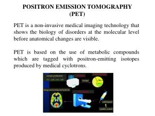
Most used tracer ? Fluor-18 – deoxyglucose = FDG Half-life-time: 110 minutes, iv injection
Imaging and measurement of glucose up-take Into normal tissue Brain, heart, liver and other organs In cancer tissue Brochial, skin, colon, liver, lymph node, esophagus, larynx-cancer in inflammation What is PET able to do with FDG ?
Visualisation of normal tissue Vessels, intestine, bone, muscle Changes in morphology anatomic variants organ-enlargement pathological changes BUT: Is not able to distinguish distinctly between malignant and benign changes What is CT able to give us ?
Combination of function PET – positron emission tomography and morphology CT – computed tomography PET/CT
Combination: Functional information from PET morphological information from CT Time saving: Reduced data acquisition time of PET/CT compared to PET alone 55 minutes vs. 20-24 minutes PET/CT: PET and CT in a single machine
FDG-PET in Oncology Diagnosis Staging Prognosis Therapy Monitoring Metabolic characterization of structural lesions Correlation with response rate and survival Responders versus Non-responders Whole-body screening
PET and lung cancer: mediastinal staging • early stage tumors • stage I (T1-2, N0) • stage II (T1-2, N1) • locally advanced tumors • stage IIIa (T1-3, N2) • stage IIIb (T1-4, N3) surgical treatment marginal resectability induction treatment non-surgical combined treatment Importance of an accurate mediastinal staging
Detection of lymph node metastases Lymph nodes pos.CTPET Sensitivity57-70 %85-92 % Specificity80-94 %90-98 % PPV76-84 %90-96 % NPV84-90 %95-98 %
T staging in NSCL with PET/CT vs. PET+CT Lardinois, Weder, Hany et al. et al., N Engl J Med 2003;348:2500-7 50 patients prospectively evaluated PET/CT additional information in 41% over PET + CT T stagePaired Sign Test P-value PET/CT vs. CT 0.001* PET/CT vs. PET <0.001* PET/CT vs. PET+CT 0.013* N stagePaired Sign Test P-Value PET/CT vs. CT 0.12 PET/CT vs. PET 0.013* ___________ PET/CT vs. PET+CT 0.021 *significant after Bonferroni correction
1 ,8 ,6 ,4 ,2 PET log-rank P = 0.008 0 0 12 24 36 48 60 Follow-up (months) = overall response = no overall response PET and lung cancer: therapy evaluation • Prognosis according to overall response 1 ,8 Cumulative survival ,6 ,4 ,2 CT log-rank P = 0.10 0 0 12 24 36 48 60 Follow-up (months)
Tumor markers in the blood FDG-PET to predict those patients who benefit from laparotomy Libutti SK et al. Ann Surg Oncol. 2001 Dec;8(10):779-86. What does PET/CT for recurrent disease? Recurrent disease PET
Liver metastases (sensitivity) ceCT 95%, PET/CT 91% Intra-hepatic recurrence after liver surgery (specificity) ceCT 50%, PET/CT 100% Local recurrence (sensitivity) ceCT 53 %, PET/CT 93 % Extra-hepatic disease (sensitivity) ceCT 64 %, PET/CT 89 % Additional findings in PET/CT changed management in 21% of cases PET/CT vs. ceCTSelzner M, Hany TF et al. Ann Surg Dec 2004
Hodgkin's disease (HD) less than 1% of all cancers 70% can be cured with combination chemotherapy and/or radiation therapy Non- Hodgkin's lymphoma (NHL)5% of all cancers less predictable than HD and has a greaterpredilection to disseminate to extra nodal sites 2 prognostic groups: low-grade NHL and aggressive NHL overall survival at 5 years is approximately 50% to 60% Lymphoma: epidemiology
Hodgkins lymphoma: FDG-PET staging • CT PET • sensitivity 79% 84% • Specificity 100% 100 % • Jerusalem et al. 2001 • Najjar et al. 2001 • Weihrauch et al. 2002
lymph node involvement sensitivity of PET/CT and ceCT was 93% and 87% specificity was 100% and 85% organ involvement sensitivity of PET/CT and ceCT was 87% and 50% specificity was 100% and 90% PET/CT lymphoma staging/restaging
HD and NHL: up to 60% with residual mass in computed tomography postive PET scan -> high probability of relaps in HD: inflammation or thymus uptake negative scan: does not exclude disease but longer DFS De Wit M et al. Ann Oncol 2001; 12:29-37 Spaepen K et al. Br J Haematol 2001; 115:272-278 Spaepen K et al. J Clin Oncol 2001; 19:414-29 Residual mass after treatment
Evaluation after first-line therapy • Time-point of re-evaluation: • after full scale chemo-therapy • Prognostic role: • significant difference: • positive predictive value for PET compared to CT regarding progression free survival (PFS) and overall survival (OS) • progression free survival (PFS) shorter for persistent uptake • Earlier evaluation? • Jerusalem G. Blood. 1999 Jul 15;94(2):429-33. 8 cycles CHOP
Time-point of re-evaluation: after 2-4 cycles of chemo-therapy Prognostic role: significant difference in PPV for PET compared to CT regarding progression PFS and OS progression free survival (PFS) shorter for persistent uptake Jerusalem G.Ann Oncol. 2003Jan;14(1):123-30. Short term follow-up: Lymphoma 4 cycles CHOP
Staging: PET/CT in 3 (15.7%) patients, ceCT in 1 patient (5.2%) Restaging: PET/CT 6 patients (14.6%), ceCT 1 patient (2.4%) Suggestion: ceCT not anymore necessary, PET/CT sufficient PET/CT findings changed treatmentSchaefer N, Hany TF Taverna et al. Radiology 2004
(PSA 82 ng/ml) Re-staging 18F-Cholin
PSA 2.1 ng/ml 10 d after surgery: 0.12 ng/ml Actually after 1 year: 0.00ng/ml Biochemical recurrence of prostate cancerSchmid DT, Hany TF. (2005) Radiology. May;235(2):623-8.