Simulation Needs in PET: Imaging Physics, Components, and System Configurations
540 likes | 619 Vues
Explore the key components, system geometries, and simulation needs in Positron Emission Tomography (PET) imaging, including source distributions, photon detection, scintillation crystals, electronics, and image reconstruction techniques. Discover the advancements in PET technology for both human and animal imaging applications.

Simulation Needs in PET: Imaging Physics, Components, and System Configurations
E N D
Presentation Transcript
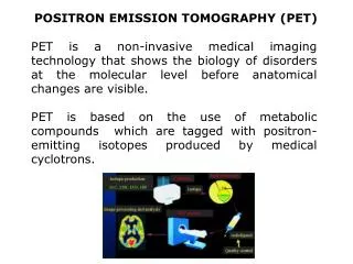
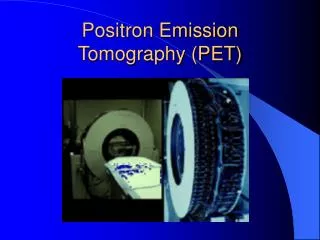
Simulation Needs in PET(Positron Emission Tomography) Chin-Tu Chen, Ph.D. Department of Radiology & Committee on Medical Physics Pritzker School of Medicine & Division of Biological Sciences The University of Chicago
PET Principle P N + e+ + n + energy E = mc2
Production of Isotopes (Mini-Cyclotron) 18O(p,n)18F
PET Isotopes 15O 13N 11C 18F 64Cu 82Rb 124I PET Tracers [15O]-O2 [15O]-H2O [15O]-H2O [15O]-CO [13N]-NH3 [18F]-FDOPA [13N]-glutamate [18F–] [11C]-acetate [18F]-FDG [11C]-palmitate [11C]-methionine
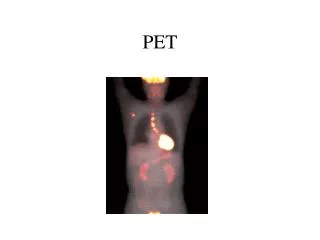
Metastatic Melanoma 71-year-old male with metastatic melanoma on left shoulder discovered 12/94. CT performed on 7/10/95 demonstrated tumor of the distal femur with negative findings in the abdomen. Bone scan on 7/13/95 showed an abnormal femur and four spine lesions. Whole-body FDG PET scan demonstrates numerous lesions throughout the body. Patient was scheduled for an amputation and total knee replacement based on CT and bone scan results. After PET found multiple lesions, surgery was cancelled, avoiding both the cost and the trauma of an operation that would not be effective. Courtesy of Amjad Ali, M.D. • Rush-Presbyterian-St. Luke’s Medical Center
Imaging of the Brain, Mind, Cognition, and Behavior
11CH3 O OCH3 N F [11C]WIN 35,428 Biochemical Imaging with Small Animals microPET
microPET Images baby rhesus monkey brain phantom (25 cc) - 1.2 mCi 18FDG 1hr. acquisition microPET EXACT HR+
Animal Studies: 11C-WIN 35,428 in collaboration with Bill Melega Vervet Monkey Rat Mouse x2 x2 Transverse Coronal
FDG Whole Body Rat Study Injected dose: 2.5 mCi Imaging time: ~2 hrs.
Mouse model with one tumor on each shoulder. The left tumor expresses the D2 receptor gene and uptakes FESP, while the tumor on the right, represses the tk gene and uptakes FPCV.
Human PET: 3-4mm; Target: 1-2mm Animal PET: 1-2 mm; Target: <0.5mm
Simulations in PET Source Distributions Imaging Physics Attenuation Scattering etc.
The neue Cologne Phantom 3 mm structure 2 mm structure
Simulations in PET Imaging System Geometry Configurations
HRRT: Octagon - 120,000 crystals 936 electronic channels4.486*109 LORs
A Benchtop Prototype for High-Throughput Animal Imaging • HRRT modules • LSO crystals with DOI capability • good spatial resolution • ~2.42mm crystal pitch • ~10mm DOI resolution • good detection sensitivity • high count rate • large detection sensitive area • ~25.2cm ×17.4cm • 72×104 crystals per layer • off-shelf, well tested, cost-effective design • adjustable energy and coincidence windows
Multi-Modality Integrative System PET/SPECT PET/MRI PET/SPECT/CT For Animal Imaging Siemens “Molecular Imaging”
Simulations in PET Photon Detection Scintillation Crystals Photon Sensors PMT/APD/SiPM
Scintillation properties of primary crystals in PET aRelative to NaI(Tl)=100 bIt is the fast scintillating component of BaF2. cx=0.1 dx=0.1
PET Components • detectors • block detectors: BGO, LSO, GSO crystals
Quadrant Sharing Design • 8x8 crystal matrix; two layer LSO LSO-fast/LSO-slow (or LSO/GSO) • 128 single crystals in 2 layers 2.1 x 2.1 x 7.5 mm3
Simulations in PET Electronics (Fast Electronic)
Custom Integrated Circuit Electronics Detector Analog ASIC Digital Coincidence ASIC Detector Digital ASIC Analog Detector Signals Processes Digital X, Y Energy, and Time Data Processes Digital Crystal Position and Time Data Processes Digital PET Coincidence Data
Serial Interface Gain Control DACs CMOS PET Front End Integrated Circuit Preamps, Variable Constant Fraction Gain Amps, Summers Discriminator* ECL Driver 6.4 mm Gated Integrators CFD Thld DAC X, Y Offset DACs 6.0 mm * U.S. Patent 5,396,187
Simulations in PET List Mode Position Energy Timing Depth-of-Interaction (DOI) Time-of-Flight (TOF)
DOI Detectors • Phoswich detectors • photo-diodes PMT GSO/LSO LSO scintillator (BGO) PMT photo diode
D Time-of-Flight Tomograph x • Can localize source along line of flight - depends on timing resolution of detectors • Time of flight information can improve signal-to-noise in images - weighted back-projection along line-of-response (LOR) x= uncertainty in position along LOR = c .t/2 Karp, et al, UPenn
5Mcts 5Mcts TOF 1Mcts TOF 1Mcts no TOF 300 ps TOF Benefit of TOF Better image quality Faster scan time 1 Mcts 5 Mcts 10 Mcts Karp, et al, UPenn
Simulations in PET Image Reconstruction Image Processing Image Analysis
Multi-Modality Bayesian Image Reconstruction Upper Two: Filtered BackProj. Lower Two: Multi-Modality Image Reconstru. Chen, Kao, et al • Co-registration of PET/SPECT with CT/MRI • Incorporation of high-resolution information from the co- • registered CT/MR images into a Bayesian image recons- • truction framework to enhance image quality of PET/SPECT • Using the co-registered CT/MR images as an anatomic map • in correction for attenuation and scatters in PET or SPECT
Dual Planar Detector High-Throughput Animal PET Imager Dual Layer D.O.I. Detectors (LSO) Variable Detector Face-to-Face Spacing
Example Reconstruction 25.2 cm 5 cm ~16 cm Noiseless data Noisy data
Simulations in PET Physiology Biochemistry Biology
Simulations in PET System Geometry/Configuration Source Distribution/Physics Photon Detection/Collection Electronic ListMode/DOI/TOF Image Reconstruction Physiological Modeling