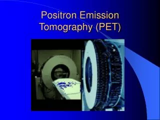
Positron Emission Tomography (PET)
420 likes | 1.62k Vues
Positron Emission Tomography (PET). History. 1975 the first commercial PET scanner was introduced 70s and 80s PET was mainly used for research 1990s being used in clinics regularly. PET vs. CT & MRI. What Can PET Detect?.

Positron Emission Tomography (PET)
E N D
Presentation Transcript
History • 1975 the first commercial PET scanner was introduced • 70s and 80s PET was mainly used for research • 1990s being used in clinics regularly
What Can PET Detect? • Coronary Artery DiseasePET imaging is unique in its ability to determine whether a patient's heart muscle will benefit from coronary artery bypass surgery.
Example: Myocardial Viability • The first heart has a mycardial infarction. The arrows point to damaged areas (‘dead’ tissue).Therefore it is assumed that the patient will not benefit from heart surgery. • The second heart is normal
TumorsPET imaging is very accurate in differentiating malignant from benign growths, as well as showing the spread of malignant tumors. PET imaging can help detect recurrent brain tumors and tumors of the lung, colon, breast, lymph nodes, skin, and other organs. Information from PET imaging can be used to determine what combination of treatment is most likely to be successful in managing a patient's tumor.
Example: Breast Cancer • The fist picture shows a malignant breast mass not shown in the conventional imaging techniques. (CT, MRI, mammogram) • Second picture is that of the same patient with enlarged left axillary lymph nodes. Through a biopsy they were found to be metastatic.
Diseases of the BrainPET imaging can provide information to pinpoint and evaluate diseases of the brain. PET imaging can show the region of the brain that is causing a patient's seizures and is useful in evaluating degenerative brain diseases such as Alzheimer's, Huntington's, and Parkinson's. Within the first few hours of a stroke, PET imaging may be useful in determining treatment therapies.
Example: Seizures • First picture is that of a normal brain. • The image of a 9 year old, female patient with a history of poorly controlled seizures. The arrow points to the area that is responsible for the seizures. Through surgical removal the seizures subsequently stop.
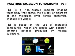
Overview of Methodology • production of positron emitting isotope in a cyclotron • chemistry of labeling compound with positron emitter and preparing compound in a form suitable for administration in humans • transport of labeled compound from chemistry group to camera group • administration (injection) of tracer compound & data acquisition with PET camera • processing of data from PET camera to extract information related to the tracer's kinetics in the body • interpretation of result
Positrons • When a positron meets an electron the collision creates two gamma rays that have the same energy but go in opposite directions. PET detects the gamma rays as they leave the patients body. The information is fed to a computer and makes a picture.
Positrons Continued… • http://www.austin.unimelb.edu.au/dept/nmpet/pet/detail/principle.html • Short clip to further illustrate the function of positrons.
Tracers • Depending on the type of tracer, different gamma rays are given off and detected. In this way multiple things can be evaluated. • Examples: ·Fluorodeoxyglucose [18F]-labeled 2-deoxyglucose (FDG) is used in neurology, cardiology and oncology to study glucose metabolism. FDG is potentially useful in differentiating benign from malignant forms of stimulated osteoblastic activity because of the high metabolic activity of many types of aggressive tumors.
·Oxygen[15O]-labeled water and oxygen are being evaluated for the quantification of myocardial oxygen consumption and oxygen extraction fraction. [15O]-labeled oxygen can also be used to measure tumor necrosis. • ·Ammonia[13N]-labeled ammonia can be used to measure blood flow. • ·Leucine[11C]-labeled methionine and leucine can be used to evaluate amino acid uptake and protein synthesis, providing an indicator of tumor viability. • ·Fluorine IonRadiolabeled fluorine ion [18F-] was once a standard agent for clinical bone scanning.
Camera Design • Many detectors are connnected to a photomultiplier tube and arranged in circular, hexagonal, or orthoganal rings. • The field of view is defined by the width of the array of detectors. • The smaller the size of detectors the better the spatial resolution of the PET system.
Interpretation of PET • Examples: case studies
Case Study • A 71 year-old male with metastatic melanoma on the left shoulder, discovered 12/94. • Original Diagnosis • A CT performed on 7/10/95 demonstrated a tumor of the distal femur and adjacent soft tissue with negative findings in the abdomen. A bone scan from 7/13/95 showed on abnormal femur and four spine lesions. • PET Findings • History • A whole-body FDG PET scan demonstrated numerous lesions throughout the body. • Change in Treatment • The patient was scheduled for an amputation based on CT and bone scan results. After the PET scan found multiple lesions, surgery was cancelled, avoiding both the cost and the trauma of an operation that would not have been effective.
Case Study 2 • History • A 64 year-old male who was found to have serum CEA elevation for 16 months after resection of colon carcinoma. • Original Diagnosis • Endoscopy with biopsy demonstrated recurrence at the site of anastamosis at the hepatic flexure. CT of the abdomen and pelvis showed no abnormality. • PET Findings • Preoperative PET scan showed a new abnormal focus in the mediastinum and two on the lumbar spine (left image), and a focus in left lobe of the liver and two in the left chest wall (right image). • Change in Treatment • The findings indicate local recurrence and metastatic disease to the liver, mediastinum, and skeleton. This patient will have to be placed in chemotherapy in addition to any surgery planned.
Case Study 3 • History • A 63 year-old male with lung cancer. A tumor was removed from the right upper lobe. • Original Diagnosis • Several months later a CT scan showed a new lesion in the left lung. • PET Findings • A whole-body FDG PET scan found focal FDG accumulation in the left lung lesion, as shown in figure 1. In addition, several other lesions unsuspected by CT were seen in the right lung and mediastinal lymph nodes. • Change in Treatment • The conventional treatment plan based on CT results would have been a thoracotomy to resect the lesion in the left lung. After the PET scan found other areas of focal FDG uptake, the treatment was switched to chemotherapy. The lesions could still be seen on a follow-up CT scan after chemotherapy, but a second PET study showed the FDG uptake had returned to normal, avoiding a possible resection.
Factors Affecting PET • Attenuation • Random Coincidences • Scatter Coincidences • Spatial Resolution • Sensitivity
Quality Control Tests • Daily Tests • Uniformity of the image is checked daily using a elictical filled with a positron emitter • Weekly Tests • Spatial Resolution is checked for by service personnel by periodically using the line spread functions • Center of Rotation is checked so that there are no deviations from the proper alignment.
Future Studies • In 2000 CTI Molecular Imaging Inc. was the first to market the combination PET/CT scanner; provides the most complete and accurate picture • PET/CT scanning allows for instantaneous results for both anatomy and function, taking the guess work out of comparison with separate images.
For More Information • www.petscaninfo.com • www.petnetpharmaceutical.com • www.nucmed.buffalo.edu • www.crump.ucla.edu