Nuclear Magnetic Resonance Spectroscopy
300 likes | 716 Vues
Nuclear Magnetic Resonance Spectroscopy. By James Buxton and Sahil Nalavade. Theory behind NMR.

Nuclear Magnetic Resonance Spectroscopy
E N D
Presentation Transcript
Nuclear Magnetic Resonance Spectroscopy By James Buxton and Sahil Nalavade
Theory behind NMR • NMR Is a technique used to determining the structure of complex biochemical molecules. It can only be used with molecules that contain atomic nuclei that have a ‘spin’. Overall spin can only be found in nuclei with have either an odd number of protons, or an odd number of neutrons or an odd number of both protons and neutrons. This is because if all the nucleons spins were paired they would cancel out. • Electrons, protons and neutrons can be thought as either spinning up or down. • A charged particle with spin creates a magnetic field along its axis (a magnetic moment). When a external magnetic field is applied the nucleus can be aligned with (lower energy) or against the field (higher energy)and theses orientations have different energies (slightly more nucleons will be in the lower state aligned with the external field). The energy difference between the higher and lower states (ΔE = hν) is so small that electromagnetic waves with low frequencies (i.e. Radio waves) can cause a nucleus to flip its orientation and absorb the wave, this region is called the radio region. • This difference in energy of the two spin states depends on which kind of nucleus it is (i.e. Different isotopes have different nucleuses) and the chemical environment that surrounds the nucleus.
Theory cont’d • A nucleon can absorb a quantum of energy in the radio region and be put into the higher spin state. When it relaxes it returns to the lower level and will emit the same radio frequency (see image) • The magnetic field experienced by the nucleus is a result of the electrons around it spinning and having a associated magnetic field . This electron generated field ensures that the field experienced by the nucleus is not the external magnetic field. This is known as Nuclear shielding and extent of it is determined by the other atoms surrounding the nucleus as the magnetic field of neighbouring atoms modifies the external magnetic field experienced by the atom within a molecule. . The amount of shielding also changes the required energy for the nucleus to change its spin. For example the frequency absorption would be minutely different for the hydrogen in –CH3 than the hydrogen –CH2-.
Theory cont’d • J-coupling (also called indirect dipole dipole coupling) is the connection between two nuclear spins due to the influence of bonding electrons on the magnetic field running between the two nuclei. J-coupling contains information about dihedral angles, which can be estimated using the Karplus equation. It is an important observable effect in 1D nuclear magnetic resonance spectroscopy.
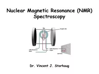
Instrumentation • A basic NMR spectrometer includes: • Very strong magnets because NMR signals are more spread out when a more intense magnetic field is used (for interpreting). • A radio transmitter coil • The sample (if it is a solid it is dissolved in a solvent that will not emit a signal such as CD2CL2 as Deuterium [D OR 2H], 16O and 12C have paired protons and neutrons which means they lack overall spin) • A radio receiver coil so that when relaxing nuclei move down to a lower energy level and emit a radio frequency it can be detected. • A computer which both analyses and records the data.
Uses of NMR • NMR is used in a wide variety of scientific disciplines for a range of purposes. • The most well known use of NMR is when used in medicine for magnetic resonance imaging (MRI).MRI works by measuring the amount of time it takes for exited protons in the human body to relax back down to the lower energy state. This time is different for all the different tissues in the body which means that a tumour can be identified as it also has a different response . In this case a computer analyses the data (in quantitative form) and gives off a coloured image (qualitative data) of the persons body that is easy for doctors to interpret. • Another very common application of NMR is in a chemical analysis. Theses can be carried out by a great number of different scientists such as forensic scientists (to identify toxins, etc) and analytical chemists (to discover/learn about/identify the structure of a specific molecule).The data comes in the form of peaks on a spectrum (quantitative data) • Other uses include non-destructive testing, In the petroleum industry to find pore size distribution and identify pore fluids (water, oil and gas) in boreholes, In quantum computing using the spin states of molecules as qubits (a unit of quantum information) as well as many other uses in a large variety of disciplines .
Reliability • It is very hard to compare frequencies from one NMR to another but, it is necessary for reliability. So, in order to standardise the measurements of different machines and under different experimental conditions, a ‘control’ sample is used as a reference. This is almost always tetramethylsilane (TMS), (CH3)4Si (see image) The reason for using TMS is because its structure ensures that all the H atoms in the molecule are in an identical environment. It is inert so it can be added to the sample without causing a reaction. Also, when looking at the data of a NMR the TMS peak is well away from the ones the experimenter would be interested in. T he difference in energy needed to change the spin state in the sample is compared to the TMS and is called ‘chemical shift’. Chemical shift is measured in ppm and its symbol is δ. The chemical shift of TMS is defined as zero. • For large molecules (for example protein molecules containing thousand of atoms ) it is likely that NMR signals will overlap, making it harder to use NMR to learn about its structure. However, some of the different forms of NMR such as solid state NMR can be used with proteins ,polymers, etc (refer to future slide).
Reliability Cont’d • NMR is very useful because it can give many types of information. It is similar to infrared spectroscopy to identify functional groups. analysis of a NMR spectrum provides information on the number and type of chemical entities in a molecule. However, NMR provides much more information than IR. However, much more information can be obtained from using NMR. • The data is fairly easy to understand for the trained eye. • Largely non destructive so it is good for expensive (DNA/RNA) or dangerous samples (as long as they are not ferromagnetic)
Reliabilitycont’d • It is considerably slower than other techniques • The atomic nucleus of an isotope in the molecule needs to have overall atomic spin. • Other limitations on sensitivity arise because NMR requires an majority of the population to be higher in the lower energy state and also that particles goes from the low energy state to the high energy state. It has been discovered that radiation and temperature can affect the atoms ability to move from energy states. However, ways have been found to decrease the sensitivity of NMR such as changing the temperature which have made them hardier.
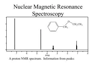
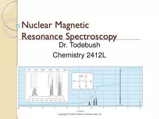
Examples of output • MRI • low resolution NMR- The chemical shifts give information about the sort of environment the hydrogen atoms are in, the ratio of the areas under the peaks gives the ratio of the numbers of hydrogen atoms in each of these environments; the number of peaks gives the number of different environments the hydrogen atoms are in. • The ratio of the number of hydrogen in the various environments can be found in the ratio of the areas under the peaks. In the case of methyl propanoate, the areas were in the ratio of 3:2:3, which is exactly what is expected for the two differently placed CH3 groups and the CH2 group. • The position of the peaks tells you useful things about what groups the various hydrogen atoms are in.. The important shifts for the groups present in methyl propanoate are:
Output cont’d • High resolution NMR – Very similar to low resolution except what were peaks on low resolution are split up into what look like many peaks in clusters. These should be treated the same way as a low resolution spectrum, just treat each cluster as a separate peak.
Proton NMR -The type of NMR that uses the differences in the magnetic properties of 1H atoms in compounds in order to identify the number of chemically distinct hydrogen ‘environments’ there are ,is known as proton NMR or 1H NMR. -When looking at the data the number of peaks is equal to the number of different bonding environments experienced by all the hydrogen nuclei in the molecule. -For example in the case of the ethyl benzene all the hydrogen in the same environment are in the same peak on the spectrum. For example all of the single hydrogen atoms that are bonded to a single carbon are together in one peak.
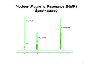
Proton NMR continued(refer to chemical shift in text and data book) • In the previous graph (because it was a high resolution NMR spectrum) the various peak s due to protons in the same environment were split into finer peaks. The amount of splitting provides information about the number of hydrogen atoms attached to adjacent carbon atoms. This interaction is known as spin-spin coupling. • The clusters are known as singlets , doublets, triplets and Quartets. • The number of peaks caused by splitting equals n+1, n is the number of H atoms on the neighbouring atom. So, A Singlet is next to carbon with no hydrogen attached • A doublet next to a CH group • A triplet next to a CH2 group • A quartet next to a CH3 group
Proton NMR cont’d • On the graph depicting ethanol(CH3CH2OH) on the next page: CH splits the signal from hydrogen attached to adjacent atoms into two peaks, CH2 splits the signal from hydrogen attached to adjacent atoms into three peaks and CH3 splits the signal from hydrogen attached to adjacent atoms into four peaks. • Also, the peak areas give us information about the amount of equivalent H atoms ( i.e. CH3 has the largest area under the peak because it has more hydrogen than the other s). • H and OH groups do not split the peaks . The reason for this is oxygen shielding . Effects of shielding • For H1 Intensity of the lines is equal to the Patterns present. • -Spectra fine structure due to spin-spin coupling is when the hydrogen atoms are on adjacent atoms ( for example H-C-C-H) and not when separated by one or more atoms (H-C-O-C-H). • To find the compound chemical shift data is also used to help to identify the environment each group was in.
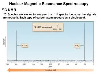
Carbon-13 NMR (refer to chemical shifts in text/data book) The second most common base isotope for NMR is Carbon-13 as most biomolecules contain carbon. 13C is naturally occurring and has nuclear spin. It is used to determine the chemical environment of carbon atoms within a molecule. In the spectra the chemical shift from the TMS is depends on the carbon atoms environment within a molecule. The shifts range from 0ppm to 200ppm (see graph below). There is only one peak produced for each carbon atom environment (see Buckyball). The proton NMR and carbon-13 NMR’s graphs are very different and should not be confused with one another.
Safety Precautions • Electrical Hazards-The NMR spectrometers in the lab operate on either 240 volts AC or 120 volts AC and have several high-voltage DC components, all of which can be • hazardous or fatal if the equipment is used improperly and accidental electrocution occurs. To prevent such accidents, safety precautions are put in place such as: • -No person may access the instruments without proper training and • Without proper authorisation. • -Extreme caution should be used whenever the instruments are being used in a way that makes it necessary to be near the console • or magnet. • -No one can access the instrument panels or spectrometer consoles unless • under the observation and guidance of the a NMR supervisor. • -Any accumulation of water around or near the instruments should be • reported to a NMR supervisor and the wet areas should be avoided • to prevent electrocution. • -Any accidental exposure to electricity must be reported to the NMR • Supervisor.
Cryogenic Liquids-Cryogenic liquids such as Liquid Nitrogen and liquid Helium are used in the NMR laboratory and both • Can be extremely dangerous. In order to prevent accidental exposure to liquid • cryogens, and to avoid asphyxiation (suffocation) in the event of a magnet quench safety precautions are put in place such: • -No person may use cryogenic liquids in the NMR laboratory without first • having been trained in the safe handling of cryogens and other dangerous substances. • -. Before using cryogenic liquids in the NMR laboratory, a NMR Facility • Supervisor (of some description)must be notified. Cryogenic liquids stored in the NMR laboratory • are not for general use. • -Protective clothing including lab coats, gloves and eye-protection will be • worn by all individuals whenever handling cryogenic liquids in the NMR • laboratory. Individuals near the vicinity of the NMR equipment when cryogens are being handled may also be required to wear • protective clothing. • -In the event of a magnet quench (the sudden evaporation of • cryogenic liquids in the magnet), all present must immediately and orderly • exit the NMR laboratory. A magnet quench is usually obvious from the • loud rushing sound of the evaporated gases escaping the magnet and may • displace sufficient Oxygen to cause asphyxiation. Since Helium is less • dense than air, exiting the laboratory by crawling on the floor is • recommended. Doors to the laboratory should be left open to aid in the • dispersal of Helium and Nitrogen gases. • -Any accidental exposure to cryogenic liquids must be reported to the • NMR supervisor in order to get the appropriate medical attention. • -
Magnetic Fields and Electromagnetic Radiation • Strong magnetic fields and several sources of electromagnetic radiation are • present in the NMR laboratory that may be dangerous. To prevent such accidents, safety precautions are put in place such as: • -Users of the NMR equipment are subjected to exposure limits to static magnetic fields. Exposure limits are the concentration of static in the workplace to which most people can be exposed without experiencing harmful effects. • -No person may enter the NMR laboratory without authorization from the • NMR supervisor. • -People with pacemakers, defibrillators, metal surgical implants or • prosthetics must stay at least 6 feet away from the magnets at all times. • -Personal articles such as hairpins or jewellery must be kept away from the • magnets at all times. • -Metal tools, carts, and gas cylinders must be kept away from the magnet • at all times.
Glass Tubes and Evacuated Storage Dewars – Glass tubes (to keep the sample in) and storage dewars (Dewar's are a container with an evacuated space between two walls that are highly reflective, capable of maintaining its contents at a near-constant temperature over relatively long periods of time-) . Like all equipment these could become quite dangerous if used incorrectly. To prevent such accidents, safety precautions are put in place such as: • -NMR Tubes must be handled with extreme caution(They are thin-walled • glass and can cause dangerous wounds. If broken) • -Never force an NMR tube into the • NMR spinner holder and never force the cap on or off an NMR tube. • -Evacuated storage dewars are in the probes on all the • spectrometers, and are sometimes used around spectrometers. • These can be very dangerous when broken as the vacuum can cause implosion. • -Always make connections to storage dewars carefully and without force. • -External storage dewars should always be wrapped in plastic mesh or tape • to prevent flying glass if they are broken. • -Broken glass should be cleaned up under the supervision of the NMR • Supervisor and should be disposed of in approved glass waste • containers. • -All injuries must be reported to the NMR supervisor. NMR Dewar
Chemical Hazards-NMR uses various hazardous chemicals (such as solvents) which can be harmful if used incorrectly, To prevent such accidents, safety precautions are put in place such as: • -NMR Solvents must be handled as specified in the Material Data Safety Sheets . • . Because there are no fume hoods in the NMR • laboratory, samples requiring a hood for safe handling must be prepared • outside of the NMR laboratory. • -Chemical spills or accidental exposure to NMR solvents must be reported • to the NMR supervisor. • -Material Data Safety Sheets should be available for all standard and calibration • samples used in the NMR laboratory.
Physical Hazards – There are many potential physical hazards in a NMR lab that can happen if the equipment is used improperly. To prevent such accidents, safety precautions are put in place such as: • -Stairways to magnets must be used with extreme caution. • -Care must be taken to avoid overturning of the magnets. No one should • ever lean on the magnets or pull on the magnets when getting up or down • from the floor (as when tuning the magnets). They are not stable and can • be overturned easily. • -Cryogenic storage dewars can also be overturned quite easily. They • should never be pulled from the top, but rather from the handles provided. • -All injuries in the NMR laboratory must be reported to the NMR supervisor.
SAMPLE QUESTION • The 1H NMR spectrum of methyl methanoate , HCOOCH3 , consists of two apparent singlets at 8.07ppm and 3.76ppm, with relative areas of 1:3. It is difficult to detect any fine structure due to spin-spin coupling because • The presence of oxygen eliminates the spin- spin coupling. • A single hydrogen atom has no interaction with a –CH3 group. • The hydrogen atoms are not on adjacent atoms. • Spin-spin coupling doe not occur in esters (Esters are derived from carboxylic acids which contains the -COOH group, and in an ester the hydrogen in this group is replaced by a hydrocarbon group of some kind).
SAMPLE QUESTION • NMR spectra can also be observed for other atoms besides from 1H. For which one of the following atoms could an NMR spectrum be observed? a)12C b)16O c) 19F d) 32S
References… • Absolute astronomy.(2009) [web page] ,www.absoluteastronomy.com/topics/Proton_NMR, date accessed 10 January 2010. • Hogendoorn, B et al.(2007) Chemistry 2 , 4TH edition. Melbourne: Heinemann. • Slade, Dr. R and Slade, M (2010) Checkpoints 2010, VCE Chemistry Unit 3. Melbourne: Cambridge University press. • Millennium House. (2008) Scientifica. NSW: Millennium House • Wikipedia. (2010) [web page] , http://en.wikipedia.org/wiki/NMR_spectroscopy, date accessed 10 January 2010. • Graham Richard, M. (2007) [webpage] michaelgr.files.wordpress.com/2007/05/nmr-pro..., date accessed 15 January 2010. • Bettelheim and March. (1991) General organic and biochemistry, 4th edition. Orlando: Saunders college publishing. • Sharp, D.W.A. ( 1984) The Penguin dictionary of chemistry. Sussex: Penguin books. • Clark, Jim. (2000) [web page] http://www.chemguide.co.uk/analysis/nmr/highres.html , date accessed 19 January 2010. • Keizer, Dr D. (2009) [WEBPAGE]http://www.bio21.unimelb.edu.au/platform-technologies/nmr-facility, data accessed 3 February 2010