Download
1 / 33
380 likes | 745 Vues
Nuclear Magnetic Resonance Spectroscopy. Dr. Todebush Chemistry 2412L. Introduction. NMR is the most powerful technique for organic structure determination Number and type of atoms in a molecule Connectivity of atoms Used to study a wide variety of nuclei: 1 H 13 C

E N D
Nuclear Magnetic Resonance Spectroscopy Dr. Todebush Chemistry 2412L
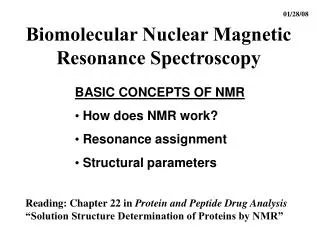
Introduction • NMR is the most powerful technique for organic structure determination • Number and type of atoms in a molecule • Connectivity of atoms • Used to study a wide variety of nuclei: • 1H • 13C • 15N, 19F, 31P • Radio-frequency radiation used to transition between energy states • 30 – 900 MHz • Transition = nuclear spin
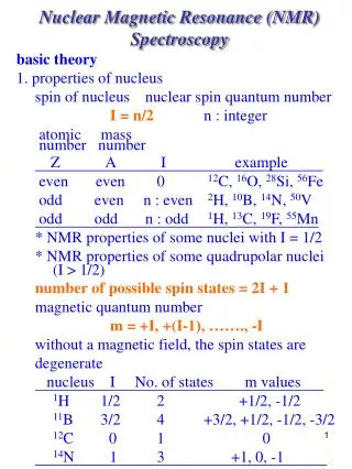
Nuclear Spin • A nucleus with an odd atomic number or an odd mass number has a nuclear spin • The spinning charged nucleus generates a magnetic field
External Magnetic Field • When placed in an external field, spinning protons act like bar magnets
Two Energy States • The magnetic fields of the spinning nuclei will align either with the external field, or against the field • A photon with the right amount of energy can be absorbed and cause the spinning proton to flip • Spin flip = resonance • Detected and recorded by the spectrometer as a signal
Magnetic Shielding • If all protons absorbed the same amount of energy in a given magnetic field, not much information could be obtained • But protons are surrounded by electrons that shield them from the external field • Circulating electrons create an induced magnetic field that opposes the external magnetic field • Effective magnetic field
Shielded Protons • Magnetic field strength must be increased for a shielded proton to flip at the same frequency • Differences detected by machine, cause differences in signals (chemical shift, d)
Protons in a Molecule • Depending on their chemical environment, protons in a molecule are shielded by different amounts • Chemically equivalent nuclei • Interchanged through bond rotation or element of symmetry • Have same absorption • Chemically different nuclei have different absorption
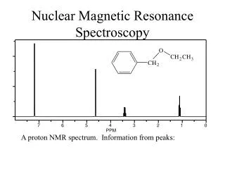
NMR Signals • The number of signals shows how many different kinds of protons are present • The location of the signals shows how shielded or deshielded the proton is • The intensity of the signal shows the number of protons of that type • Signal splitting shows the number of protons on adjacent atoms
Tetramethylsilane • TMS is added to the sample • Since silicon is less electronegative than carbon, TMS protons are highly shielded • Signal defined as zero • Organic protons absorb downfield (to the left) of the TMS signal • Deuterated solvent signal
Chemical Shift • Measured in parts per million • Ratio of shift downfield from TMS (Hz) to total spectrometer frequency (Hz) • Same value for 60, 100, or 300 MHz machine • Called the delta (d) scale
Delta Scale downfield upfield
Location of Signals • More electronegative atoms deshield more and give larger shift values (downfield) • Effect decreases with distance • Additional electronegative atoms cause increase in chemical shift
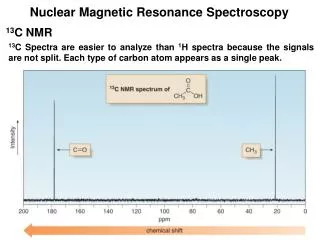
13C-NMR • 12C has no magnetic spin • 13C has a magnetic spin, but is only 1% of the carbon in a sample • Signals are weak, get lost in noise • Hundreds of spectra are taken, averaged • Signal = one sharp line for each different type of carbon
3-Pentanone • How many signals? • Chemical shifts: • sp3 C upfield • sp, sp2 C downfield • C adjacent to en atom downfield
2-Butanone • How many signals? • Chemical shifts?
How is 13C useful for reactions we have studied? • Zaitsev vs. non-Zaitsev 7 signals 5 signals
1H-NMR • More info than 13C-NMR • Given a structure, how many signals are expected? • How many sets of H in each molecule? • Isomers • Same molecular formula • Same IR stretches • Different NMR
Chemical shifts in 1H-NMR • Info about type of H giving rise to signal • Strongly shielded = upfield (to the right) • Less shielded = downfield (to the left) • Most common shifts: • Wade Appendix 1A • Wade, Table 13-3 • -CH2-O-C(O)- ranged from 3.7-4.7 ppm
O-H and N-H Signals • Chemical shift depends on concentration • Hydrogen bonding in concentrated solutions deshield the protons, so signal is around 3.5 for N-H and 4.5 for O-H
Using chemical shifts • Given a structure, predict d • Use to distinguish between two structures • Example: • Constitutional isomers • Each with 2 sets of H’s
Intensity of Signals • The area under each peak is proportional to the number of protons • Shown by integration line • Height a area under peak a # H’s in set • Measure height with ruler or look at graph paper • Ratio of height = ratio of hydrogens