Download
1 / 23
260 likes | 709 Vues
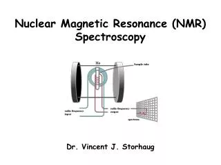
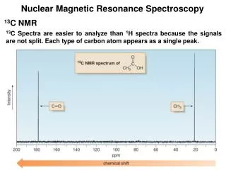
01/28/08. Biomolecular Nuclear Magnetic Resonance Spectroscopy. BASIC CONCEPTS OF NMR How does NMR work? Resonance assignment Structural parameters. Reading: Chapter 22 in Protein and Peptide Drug Analysis “Solution Structure Determination of Proteins by NMR”. Nuclear Spin.

E N D
01/28/08 Biomolecular Nuclear Magnetic Resonance Spectroscopy BASIC CONCEPTS OF NMR • How does NMR work? • Resonance assignment • Structural parameters Reading: Chapter 22 in Protein and Peptide Drug Analysis “Solution Structure Determination of Proteins by NMR”
Nuclear Spin • Nuclear spin angular momentum is a quantized property of the nucleus in each atom, which arises from the sub-atomic properties of neutrons and protons • The nuclear spin angular momentum of each atom is represented by a nuclear spin quantum number (I) • All nuclei with even mass numbers have I=0,1,2… • All nuclei with odd mass numbers have I=1/2,3/2... • NMR is possible with all nuclei except I=0, but I=1/2 has simplest physics Biomolecular NMR primarily1H, 13C, 15N (31P)
Efficiency factor- nucleus DE = h g Bo Constants Strength of magnet Spin 1/2 Nuclei in a Magnetic Field Bo Energy
Intrinsic Sensitivity of Nuclei Nucleusg % Natural Relative Abundance Sensitivity 1H 2.7 x 10⁸ 99.98 1.0 13C 6.7 x 10⁷ 1.11 0.004 15N -2.7 x 10⁷ 0.36 0.0004 31P 1.1 x 10⁸ 100 0.5 Prepare samples enriched in these nuclei
Sensitivity (S) ~ D pop. (N vs N) Efficiency factor- nucleus N N -DE/kT = e S ~ DN = DE = h g Ho Constants Strength of magnet Variables Affecting Sensitivity - DE is very small DN is small - DN ~ 1:105 (at room T) NMR has low sensitivity requires lots of sample! Increase sensitivity by increasing magnetic field strength or reducing electronic noise (cryo-probes)
Bo DE Equilibrium B1 Pump in energy (RF transmitter) hn = DE Non-equilibrium NMR signals Release energy (RF receiver) hn = DE Equilibrium The Resonance Experiment Strength of signal D (population)
NMR TerminologyChemical Shift & Linewidth The exact resonance frequency (chemical shift) is determined by the electronic environment of the nucleus
Scalar and Dipolar Coupling Through Space Through Bonds • Coupling of nuclei gives information on structure
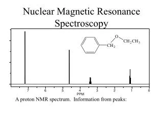
OH CH2 CH3 The key attribute: use the scalar and dipolar couplings to match the set of signals with the molecular structure Resonance Assignment CH3-CH2-OH Which signal from which H atoms?
F1 F2 2D NMR Spectra FacilitateIdentification of Coupling Coupled spins
Biomolecules Have Many Signals 1H NMR Spectrum of Ubiquitin ~75 residues, ~500 1H resonances • Terminology: signals are overlapped
Challenges For Using NMR to Study Biological Macromolecules • Hundreds-thousands of signals! • Must assign the specific signal for each atom • Thousands of couplings between nuclei- these also need to be assigned
Critical Features of Protein NMR Spectra • Regions of the spectrum correspond to different parts of the amino acid • Tertiary structure leads to increased dispersion of resonances
Regions of the 1H NMR Spectrumand Dispersion by the 3D Fold What would the unfolded protein look like?
Critical Features of NMR Spectra of Biomolecules • Regions of the spectrum correspond to different parts of the amino acid • Tertiary structure leads to increased dispersion of resonances • Bio-macromolecules are polymers The nuclei are coupled to some (but not all!) other nuclei
Spectra of Biomacromolecules:Overlapped Sub-Spectra *Groups of coupled nuclei* Each residue in the sequence gives rise to an independent NMR sub-spectrum, which is much simpler than the complete spectrum Methods have been developed to extract each sub-spectrum from the whole
T L R S G G S Strategy to AssignResonances in a Protein • Identify resonances for each residue (scalar) • Put amino acids in order (dipolar) 1 2 3 4 5 6 7 R - G - S - T - L - G - S Same idea for any biopolymer (e.g. DNA, RNA)
Even Sub-Spectra are Overlapped! 1H NMR Spectrum of Ubiquitin • Resolve resonances by multi-dimensional experiments
Use 2D NMR to Resolve Overlapping Signals Sub-spectra overlapped 1D Crosspeaks resolved! 2D Coupled spins
Hb Ha HN Multi-Dimensional NMR If 2D cross peaks overlap go to 3D or 4D …..
t2 t1 t3 Another Solution tothe Overlap Challenge • Increase dimensionality of spectra to better resolve signals: 1D2D3D4D…. • Use attached nuclei (13C,15N) to distinguish Hz HA
Multi-Dimensional Heteronuclear NMR 15N-1H HSQC F1 Chemical Shift (15N) F2 Chemical Shift (1H)
Advantages ofHeteronuclear nD NMR Uses a second nucleus to resolve overlap of the first: chemical shift of each nucleus is characteristic/sensitive to different factors More information to identify resonances Less sensitive to MW because this strategy uses large 1 and 2-bond scalar couplings