Nuclear Magnetic Resonance Spectroscopy
360 likes | 555 Vues
Nuclear Magnetic Resonance Spectroscopy. Renee Y. Becker Valencia Community College CHM 2011C. The Use of NMR Spectroscopy. Used to determine relative location of atoms within a molecule Most helpful spectroscopic technique in organic chemistry

Nuclear Magnetic Resonance Spectroscopy
E N D
Presentation Transcript
Nuclear Magnetic Resonance Spectroscopy Renee Y. Becker Valencia Community College CHM 2011C
The Use of NMR Spectroscopy • Used to determine relative location of atoms within a molecule • Most helpful spectroscopic technique in organic chemistry • Related to MRI in medicine (Magnetic Resonance Imaging) • Maps carbon-hydrogen framework of molecules • Depends on very strong magnetic fields
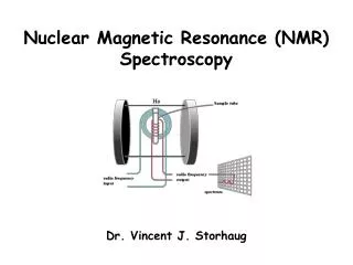
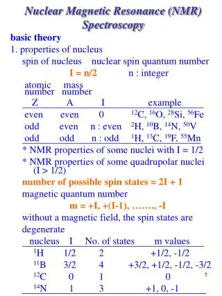
Nuclear Magnetic Resonance Spectroscopy • 1H or 13C nucleus spins and the internal magnetic field aligns parallel to or against an aligned external magnetic field (See Figure 13.1) • Applying an external magnetic field, Bo, the proton or nucleus will orient parallel or anti-parallel to the orientation of the external field. • The parallel orientation of the proton or nucleus is lower in energy than the anti-parallel orientation. • Radio energy of exactly correct frequency (resonance) causes nuclei to flip into anti-parallel state • Energy needed is related to molecular environment (proportional to field strength, Bo) – see Figure 13.2
Nuclear Magnetic Resonance Spectroscopy (1H) • The energy of the radiation required is within the radio frequency range. • The energy required is dependent upon the nucleus and the strength of the magnetic field. • A proton in a magnetic field of 1.41 telsa requires a E.M. radiation of 60 MHz to resonate. • E = 2.4 x 10⁻⁵ kJ/mol • I.R. energies 48 kJ/mol
The Nature of NMR Absorptions • Electrons in bonds shield nuclei from magnetic field • Different signals appear for nuclei in different environments
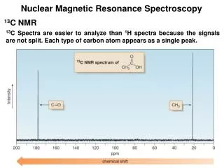
1H 13C
The NMR Measurement • The sample is dissolved in a solvent that does not have a signal itself and placed in a long thin tube • The tube is placed within the gap of a magnet and spun • Radiofrequency energy is transmitted and absorption is detected • Species that interconvert give an averaged signal that can be analyzed to find the rate of conversion
Chemical Shifts • The relative energy of resonance of a particular nucleus resulting from its local environment is called chemical shift • NMR spectra show applied field strength increasing from left to right • Left part is downfield right partis upfield • Nuclei that absorb on upfield side are strongly shielded. • Chart calibrated versus a reference point, set as 0, tetramethylsilane [TMS]
Chemical Shifts • Let’s consider the just the proton (1H) NMR. • 60 MHz NMR experiments are carried out with a constant RF of 60 MHz and the magnetic field is varied. When a spin-flip occurs (resonance), it is detected by an R.F. receiver.
Measuring Chemical Shift • Numeric value of chemical shift: difference between strength of magnetic field at which the observed nucleus resonates and field strength for resonance of a reference • Difference is very small but can be accurately measured • Taken as a ratio to the total field and multiplied by 106 so the shift is in parts per million (ppm) • Absorptions normally occur downfield of TMS, to the left on the chart
Measuring Chemical Shift Remember: the chemical shift is in ppm.
1H NMR Spectroscopy and Proton Equivalence • Proton NMR is much more sensitive than 13C and the active nucleus (1H) is nearly 100 % of the natural abundance • Shows how many kinds of nonequivalent hydrogens are in a compound • Equivalent H’s have the same signal while nonequivalent are different • There are degrees of nonequivalence
Chemical Shifts in 1H NMR Spectroscopy • Lower field signals are H’s attached to sp2C • Higher field signals are H’s attached to sp3C • Electronegative atoms attached to adjacent C cause downfield shift (deshielding) • Decreases electron density around protons • See Tables 13-2 and 13-3 for a complete list
The most important perturbation of the NMR frequency for applications of NMR is the 'shielding' effect of the surrounding electrons. In general, this electronic shielding reduces the magnetic field at the nucleus (which is what determines the NMR frequency).
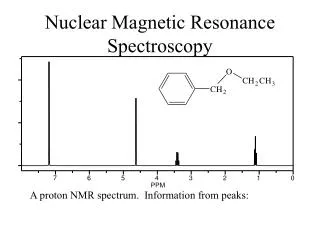
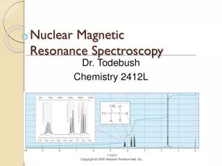
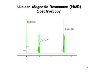
Integration of 1H NMR Absorptions: Proton Counting • The relative intensity of a signal (integrated area) is proportional to the number of protons causing the signal • This information is used to deduce the structure • For example in ethanol (CH3CH2OH), the signals have the integrated ratio 3:2:1 • For narrow peaks, the heights are the same as the areas and can be measured with a ruler
Integration of 1H NMR Absorptions: Proton Counting • This is proportional to the relative number of protons causing each signal. • An integration ratio of 1.5:1 is consistent with a 6:4 ratio of protons as with a 3:2 ratio of protons. • How many signals would you expect from the 1H NMR spectrum of chloromethyl methyl ether, ClCH2OCH3, and what would you expect the signal area ratios to be?
Spin-Spin Splitting in 1H NMR Spectra • Peaks are often split into multiple peaks due to interactions between nonequivalent protons on adjacent carbons, called spin-spin splitting • The splitting is into one more peak than the number of H’s on the adjacent carbon (“n+1 rule”) • The set of peaks is a multiplet (2 = doublet, 3 = triplet, 4 = quartet)
Rules for Spin-Spin Splitting • Equivalent protons do not split each other • The signal of a proton with n equivalent neighboring H’s is split into n + 1 peaks • Protons that are farther than two carbon atoms apart do not split each other • Unless a pi system can be used to tunnel
13.12 More Complex Spin-Spin Splitting Patterns • Spectra can be more complex due to overlapping signals, multiple nonequivalence • Example: trans-cinnamaldehyde
Analysis of NMR Spectra • The NMR spectra provides the following information that can assist in the determination of chemical structure • The number of signals • The chemical shift • The intensity of the signal (area under each peak) • The splitting of each signal