Download
1 / 23
240 likes | 435 Vues
Bi/CNS 150 Lecture 7 Monday October 14, 2013 Structure & function of glutamate receptors Henry Lester. Chapter 10 (211-227). Superfamilies of ligand-gated ion channels that are synaptic receptors. A. ACh, Serotonin 5-HT3, GABA, (invert. GluCl, dopamine, tyrosine) receptor-channels.

E N D
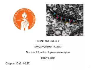
Bi/CNS 150 Lecture 7 Monday October 14, 2013 Structure & function of glutamate receptors Henry Lester Chapter 10 (211-227)
Superfamilies of ligand-gated ion channels that are synaptic receptors • A. ACh, Serotonin 5-HT3, GABA, (invert. GluCl, dopamine, tyrosine) receptor-channels Most ^ Modified from Figure 10-7
The Hippocampus: a favorite model system for neuroscience The “tri-synaptic pathway” See also Chapter 67
400 nm Electron micrograph of hippocampal synapse “Map” of micrograph to the left
A postsynaptic density, with a cartoon of important proteins
Selectivity Filter Ionotropic Glutamate Receptors:3 transmembrane helices plus a selectivity filter per subunit x 4 subunits Figure 10-8 There are also G protein-coupled glutamate receptors
3 families of Ionotropic Glutamate Receptorsare named by their selective synthetic agonists Family Subunits AMPA GluR1-4 (Most are GluR1/2 or GluR2/3) Kainate GluR5-7 NMDA NR1* (*obligatory) NR2A or B or C or D
AMPA and Kainate Receptors: With GluR2 subunit: • permeable only to K+ and Na+ Without GluR2 subunit: • Ca2+-permeable (and K+, Na+) NMDA Receptors: • Permeable to K+, Na+, Ca2+ Ion Selectivity of Glutamate Receptor Channels
RNA Editing Determines Ca2+Permeability of AMPA Receptors #1 Q= R= Transcribed codon: CAG Edited codon: CIG Right: Modified from Zigmond et al. (Eds.) Fundamental Neuroscience, Sinauer (1999)
RNA Editing Determines Ca2+ Permeability of AMPA Receptors #2 Fig. 10-9
Isolating AMPA-Receptor and NMDA-Receptor CurrentsWith Selective Blockers #1 There is outward NMDA receptor current Nestler, Hyman, & Malenka, Molecular Neuropharmacology
Isolating AMPA-Receptor and NMDA-Receptor CurrentsWith Selective Blockers #2 blocks AMPA receptors There is **no** inward NMDA receptor current blocks NMDA receptors Nestler, Hyman, & Malenka, Molecular Neuropharmacology
AMPA response is faster than NMDA response Figure 10-6
NMDA Receptors • Are composed of NR1 subunits and four different NR2 subunits; NR2A, NR2B, NR2C, and NR2D. • They contain two NR1 subunits, and a pair of NR2 subunits, which can be identical or mixed. • NR1 subunits are similar in size to GluR1-4; they are necessary to form the receptor channel, and they bind the co-ligands glycine or d-serine. • NR2 subunits are approximately twice as long as NR1 subunits. They bind glutamate, and their very long cytosolic tails bind signal transduction molecules and link the receptors to the postsynaptic density scaffold.
From Lecture 1 Time required to exchange waters of hydration
In NMDA receptors, the selectivity filter also serves as a Mg 2+ binding site, producing a “coincidence detector”. Their channel opens only when two events happen concurrently: 1. Binding of glutamate • 2. Strong postsynaptic membrane depolarization • (as by an action potential) • The depolarization relieves block by Mg2+ Modified from Zigmond et al. (Eds.) Fundamental Neuroscience, Sinauer (1999) Greater detail on learning & memory in a later lecture The coincidence readout: NMDA receptors are very permeable to Ca2+
Behavior of the NMDA receptor current Channels are blocked by Mg2+ at negative potentials Singlechannels Macroscopic I-V relations Figure 10-5
Synthetic fluorescent dyes such as Fura-2 Detect intracellular Ca2+ transients Fura-2 “Ratio Imaging” From Grynkiewicz, Poenie, and Tsien (1985) J. Biol. Chem. 260, 3440.
(repeated glutamate pulses) NMDA Receptors Mediate Synaptic Ca2+ Entry Lisman et al. Nature Rev. Neurosci.3: 175 (2002)
In green fluorescent protein (GFP), the fluorophore is well protected from the environment, by a “can” of β strands. If we render this protection sensitive to a olecule or condition, we have a good fluorescent sensor. .
Genetically encoded Ca2+ sensors Circularly permuted enhanced green fluorescent protein Calmodulin Akerboom, (30 other authors), Looger, J Neurosci 2012
Control of Synaptic Plasticity by NMDA Receptors (thought to underlie some aspects of memory and learning) Greater detail in a later lecture • The central role of Ca2+ in initiation of long-term plastic changes • The “Ca2+ hypothesis” for control of synaptic plasticity • Measurement of cytosolic Ca2+ with fluorescent dyes. • Control of postsynaptic Ca2+ by “spike timing” • Structure and behavior of the NMDA receptor • Subunit composition • Gating (“coincidence detection”) • Ion selectivity (Na+, K+, Ca2+) • Kinetics, NMDA receptors are slower than AMPA receptors • Pharmacology • The NMDA receptor is also a “scaffold.” • The postsynaptic density • LTP and LTD are triggered by Ca2+-sensitive signaling machinery located near the mouth of the NMDA receptor. • Critical components of the postsynaptic density • Biochemical pathways mediating changes in synaptic strength
Henry Lester’s office hours Mon, Fri 1:15 – 2 Red Door End of Lecture 7