Download
1 / 22
220 likes | 377 Vues
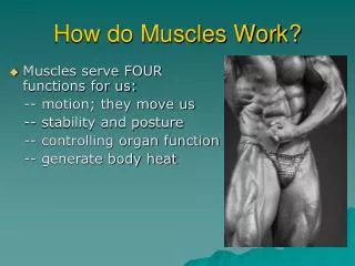
How muscles work. Skeletal Muscle Structure. Components of a Muscle Fiber. The Sarcomere. Dfdfsdfsd ssdfdf. Gfgdf bvbcv. Gfgdf bvbcv. Gfgdf bvbcv. Cvcx. Z-line. Sliding filament theory. Fig. 10-9. Overview of the process. Fig. 10-9. Overview of the process.

E N D
The Sarcomere Dfdfsdfsd ssdfdf Gfgdf bvbcv Gfgdf bvbcv Gfgdf bvbcv Cvcx Z-line
Fig. 10-9. Overview of the process The muscle fiber is stimulated.
Fig. 10-9. Overview of the process The muscle fiber is stimulated. Ca2+ ions are released.
Fig. 10-11. “End-on” view of thick & thin filaments, showing the effect of calcium ions after release from the S.R.
Fig. 10-9. Overview of the process The muscle fiber is stimulated. Ca2+ ions are released.
Fig. 10-9. Overview of the process The muscle fiber is stimulated. Ca2+ ions are released. Thin filaments move to middle of sarcomere.
Fig. 10-12 Calcium attaches to troponin/ tropomyosin; they roll away, exposing the active site on actin.
Fig. 10-12 After attachment, the cross-bridges pivot, pulling the thin filaments. Myosin cross-bridges attach to active site on actin.
Energy from the splitting of the fresh ATP allows repositioning of the myosin head. A fresh ATP replaces the ADP+Pi, allowing myosin and actin to detach.
Fig. 10-12 This leads back to Step 1, which continues the cycle as long as calcium ions are attached to troponin/tropomyosin.
Fig. 10-9. Overview of the process The muscle fiber is stimulated. Ca2+ ions are released. Thin filaments move to middle of sarcomere.
Fig. 10-9. Overview of the process The muscle fiber is stimulated. Ca2+ ions are released. Thin filaments move to middle of sarcomere. Muscle fiber contracts.
Fig. 10-9. Overview of the process The muscle fiber is stimulated. Ca2+ ions are released. Thin filaments move to middle of sarcomere. Muscle fiber contracts. Muscle tension increases.