Download
1 / 106
1.13k likes | 1.33k Vues
Learn about the anatomy of the forearm and elbow, with a focus on the extensor muscles in the posterior compartment. Discover the functions, origins, insertions, actions, and innervations of key muscles like Extensor Carpi Radialis Longus and Brachioradialis.

E N D
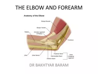
Anatomy of The Forearm And Ellbow Dr. Fadel Naim Orthopedic Surgeon IUG
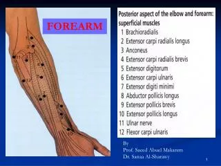
Extensor Muscles of the Forearm • In the posterior (extensor-supinator) compartment of the forearm • All are innervated by the radial nerve • Three functional groups: • Muscles that extend and abduct or adduct the hand at the wrist joint • Extensor carpi radialis longus • Extensor carpi radialis brevis • Extensor carpi ulnaris • Muscles that extend the medial four digits • Extensor digitorum • Extensor indicis • Extensor digiti minimi • Muscles that extend or abduct the thumb • Abductor pollicis longus [APL] • Extensor pollicis brevis [EPB] • Extensor pollicis longus [EPL]
Posterior compartment: • Superficial group • Extensorcarpi radialis brevis • Extensor digitorum • Extensor digiti minimi • Extensor carpi ulnaris • Anconeus muscle Attached by a common extensor tendon to the lateral epicondyle • Posterior compartment: • Deepgroup • Supinator • Abductor pollicis longus • Extensor pollicis brevis • Extensor indicis
Brachioradialis • Origin • Proximal two-thirds of lateral supracondylar ridge of humerus • Insertion • Lateral surface of distal end of radius • Action • Flexes forearm • Innervation • Radial nerve (C5, C6, and C7)
Brachioradialis • This fusiform muscle forms the lateral border of the cubital fossa • Exceptional among muscles of the "posterior" (extensor- supinator) compartment in that • It flexes the forearm at the elbow • Especially when quick movement is required and when a weight is lifted during slow flexion of the forearm. • The brachioradialis and the supinator are the only muscles of the compartment that do not cross the wrist. • To test the brachioradialis: • The elbow joint is flexed against resistance with the forearm in the midprone position. • If acting normally, the muscle can be seen and palpated.
Anconeus • Origin • lateral epicondyle of humerus • Insertion • olecranon and superior portion of shaft of ulna. • Action • Extension forearm at elbow joint • Nerve supply • radial nerve
Extensor Carpi Radialis Longus • Origin • Lateral supracondyle ridge of humerus • Insertion • Base of 2rd metacarpal • Action • Extend and abduct hand at wrist joint • Innervation • Radial nerve (C6 and C7)
Extensor Carpi Radialis Longus • To test the extensor carpi radialis longus • The wrist is extended and abducted with the forearm pronated. • If acting normally, • The muscle can be palpated infero-posterior to the lateral side of the elbow. • Its tendon can be palpated proximal to the wrist.
Extensor Carpi Radialis Brevis • Origin • Lateral epicondyle of humerus • Insertion • Base of 3rd metacarpal • Action • Extend and abduct hand at wrist joint • Innervation • Deep branch of radial nerve (C7 and C8)
The extensor carpi radialis brevis and longus act together to steady the wrist during flexion of the medial four digits.
Extensor Digitorum • Origin • Lateral epicondyle of humerus • Insertion • Extensor expansions of medial four digits • Action • Extends medial four digits at metacarpophalangeal joints;Extends hand at wrist joint • Innervation • Posterior interosseous nerve (C7 and C8), the continuation of the deep branch of the radial nerve
Extensor Digitorum • The principal extensor of the medial four digits • Its four tendons proximal pass through a common synovial sheath, deep to the extensor retinaculum with the tendon of the extensor indicis • Adjacent tendons are linked by intertendinous connections. • Because of presence of the intertendinous connections extension of one finger is impossible • The index finger has greater freedom because its tendon is not connected to the other tendons
Extensor Digitorum • To test the extensor digitorum: • The forearm is pronated and the fingers are extended. • The person attempts to keep the fingers extended at the metacarpo-phalangeal joints as the examiner exerts pressure on the proximal phalanges by attempting to flex them • If acting normally, the extensor digitorum can be palpated in the forearm, and its tendons can be seen and palpated on the dorsum of the hand.
Extensor Digiti Minimi • Origin • Lateral epicondyle of humerus • Insertion • Extensor expansion of 5th digit • Action • Extends 5th digit at metacarpophalangeal and interphalangeal joints • Innervation • Posterior interosseous nerve (C7 and C8), the continuation of the deep branch of the radial nerve
Extensor Digiti Minimi • This fusiform slip of muscle is a partially detached part of the extensor digitorum. • The tendon of this extensor of the little finger runs through a separate compartment deep to the extensor retinaculum • Then divides into two slips; • The lateral one is joined to the tendon of the extensor digitorum
Extensor Carpi Ulnaris • Origin • Lateral epicondyle of humerus and posterior border of ulna • Insertion • Base of 5th metacarpal • Action • Extends and adducts hand at wrist joint • Innervation • Posterior interosseous nerve (C7 and C8), the continuation of the deep branch of the radial nerve
Extensor Carpi Ulnaris • Distally, its tendon runs in a groove between the ulnar head and the styloid process • Extends and adducts the hand at the wrist joint simultaneously when acting independently. • Acting with the extensor carpi radialis, it extends the hand • Acting with the flexor carpi ulnaris, it adducts the hand. • Like the extensor carpi radialis longus, it is indispensable when making the fist. • To test the extensor carpi ulnaris: • The forearm is pronated and the fingers are extended. The extended wrist is then adducted against resistance. • If acting normally: • The muscle can be seen and palpated in the proximal part of the forearm • The tendon can be felt proximal to the head of the ulna.
Supinator • Origin • Lateral epicondyle of humerus • radial collateral and annular ligaments • supinator fossa and crest of ulna • Insertion • surface of proximal 1/3 of radius • Lateral • Posterior • Anterior • Action • Supinates forearm • Innervation • Deep branch of radial nerve (C5 and C6)
Abductor pollicis Longus • Origin: • Posterior surfaces of • Ulna • Radius • Interosseous membrane • Insertion: • Base of 1st metacarpal • Action: • Abducts thumb • Extends it at carpometacarpal joint • Innervation: • Posterior interosseous nerve (C7 and C8), the continuation of deep branch of radial nerve
Abductor Pollicis Longus • It acts with the abductor pollicisbrevis during abduction of the thumb • With the extensor pollicis during extension of this digit. • To test the APL: • The thumb is abducted against resistance at the metacarpophalangeal joint. • If acting normally the tendon of the muscle can be seen and palpated • At the lateral side of the snuffbox and on the lateral side of the adjacent EPB tendon.
Extensor Pollicis Brevis • Origin • Posterior sufraces of radius and interosseous membrane • Insertion • Base of proximal phalanx of thumb • Action • Extends proximal phalanx of thumb at carpometacarpal joint • Innervation • Posterior interosseous nerve (C7 and C8), the continuation of the deep branch of the radial nerve
Extensor Pollicis Longus • Origin • Posterior surface of middle 1/3 of ulna and interosseous membrane • Insertion • Base of distal phalanx of thumb • Action • Extends distal phalanx of thumb at carpometacarpal and interphalangeal joints • Adducts the extended thumb and rotates it laterally • Innervation • Posterior interosseous nerve (C7 and C8), the continuation of the deep branch of the radial nerve
Extensor Pollicis Longus • The tendon of the EPL passes medial to the dorsal tubercle of the radius, using it as a trochlea changing its line of pull as it proceeds to the base of the distal phalanx of the thumb.. • To test the EPL: • The thumb is extended against resistance at the interphalangeal Joint. • If acting normally, the tendon of the muscle can be seen and palpated on the medial side of the anatomical snuff box
Extensor Indicis • Origin • Posterior sufrace of ulna • interosseous membrane • Insertion • Extensor expansion of 2nd digit • Action • Extends 2nd digit and helps to extend hand • Innervation • Posterior interosseous nerve (C7 and C8), the continuation of the deep branch of the radial nerve
Arteries of Forearm • 1 brachial • 2 radial • 3 radial recurrent • 4 superficial radial • 5 deep radial • 6 ulnar • 7 anterior ulnar recurrent • 7 posterior ulnar recurrent • 8 common interosseous • 9 posterior interosseous • 10 anterior interosseous • 11 superficial branch • 12 deep branch • In the hand, the ulnar and radial arteries anastomose, formiug the superficial and deep palmar arterial arches.
Ulnar Artery • The larger of the two terminal branches of the brachial artery • Begins in the cubital fossa just medial to the biceps tendon • Descends through the anterior compartment of the forearm, deep to the pronator teres. • Then passes distally over the anterior aspect of the wrist to the palm.
Ulnar Artery • Pulsations of the artery can be palpated on the lateral side of the flexor carpiulnaris tendon, where it lies anterior to the ulnar head. • The ulnar nerve is on the medial side of the ulnar artery
Branches of the Ulnar Artery in the Forearm • Anterior and posterior ulnar recurrent • Just distal to elbow joint • Anterior ulnar recurrent artery passes superiorly • Posterior ulnar collateral artery passes posteriorly • Anastomose with ulnar collateral and interosseous recurrent arteries
Branches of the Ulnar Artery in the Forearm • Common interosseous artery: • Just distal to bifurcation of brachial artery • After a short course, terminates by dividing into anterior and posterior interosseous arteries • Anterior and posterior interosseous arteries: • Pass to anterior and posterior sides of interosseous membrane • Anteriorinterosseous artery supplies both anterior and posterior compartmentsin distal forearm • The posterior interosseous artery gives off the recurrent interosseous artery • Participates in the arterial anastomoses around the elbow
Branches of the Ulnar Artery in the Forearm • Dorsal and palmar carpal branches • Anastomose with corresponding branches of radial artery to form dorsal and palmar carpal arches, providing collateral circulation at wrist
Radial Artery • Smaller than the ulnar artery • Begins in the cubitalfossa near the neck of the radius • It passes inferolaterallydeep to the brachioradialis • The course of the radial artery in the forearm is represented by a line joining the midpoint of the cubitalfossa to a point just medial to the radial styloid process. • The radial artery leaves the forearm by winding around the lateral aspect of the wrist and crosses the floor of the anatomical snuff box
The Branches Of The Radial Artery • Radial recurrent artery • Lateral side of radial artery, just distal to its origin • Ascends on supinator and then passes between brachioradialis and brachialis • Dorsal and palmar Radial arteryat level of wrist • Anastomose with corresponding branches of ulnar artery to form dorsal and palmar carpal arches
Median Nerve • The median nerve is the principal nerve of the anterior compartment of the forearm • It enters the forearm with the brachial artery and lies on its medial side. • It leaves the cubital fossa by passing between the heads of the pronator teres • The nerve then passes deep to the FDS and continues distally through the middle of the forearm, between the FDS and the FDP.
Near the wrist, the median nerve becomes superficial by passing between the tendons of FDS and flexor carpi radialis, deep to the palmaris longus tendon
Branches of the Median Nerve • No branches in the arm, other than small twigs to the brachial artery • The branches of the median nerve arise in the forearm and hand as follows: • Articular branches to the elbow joint • Muscular branches supply: • The pronator teres • Pronator quadratus • All the flexor muscles • Except the flexor carpi uluaris and the medial half of the FDP.
The anterior interosseous nerve • The anterior interosseous nerve • Arises from the median nerve in the distal part of the cubital fossa • Passes distally on the interosseous membrane with the anterior interosseous branch of the ulnar artery. • It runs between the FDP and flexor pollicis longus to reach the pronator quadratus. • It supplies these muscles • The FDP • {lateral half sending tendons to digits 2 and 3) } • Flexor pollicis longus • Pronator quadratus • Sends articular branches to the wrist joint.
The recurrent branch of the median nerve (C8 and T1): • Arises from the median nerve as soon as it passes distal to the flexor retinaculum. • It loops around the distal border of this retinaculum to supply the thenar muscles.
Palmar coetaneous branch of the median nerve. The course of nerve may vary in 4 important ways • Normally, the nerve branches off 5 cm proximal to the wrist. It runs along the ulnar side of the tendon of the flexor carpi radialis before crossing the flexor retinaculum. • The nerve divides into two major branches, medial and lateral, while crossing the flexor retinaculum to supply the skin of the thenar eminence • Less often, the nerve arises from the median nerve in two distinct branches, which travel separately across the wrist. • The nerve may arise within the carpal tunnel and penetrate the flexor retinaculum to supply the skin of the thenar eminence. • The nerve may be absent, replaced by a branch derived from the radial nerve, the musculocutaneous nerve, or the ulnar nerve.
At the distal border of the flexor retinaculum, the median nerve divides into two branches • The medial branch: • sends cutaneous branches to the adjacent sides of the ring and middle fingers, and to the adjacent sides of the middle and index fingers. • The lateral branch • sends cutaneous branches to the radial side of the index finger and to both sides of the thumb.
Ulnar Nerve • It passes posterior to the medial epicondyle of the humerus,
Ulnar Nerve • The ulnar nerve enters the forearm bypassing between the heads of the flexor carpiulnaris
Ulnar Nerve • It then passes inferiorly between the flexor carpiulnaris and FDP, • supplying the ulnar (medial) part of the muscle that sends tendons to digits 4 and 5. • The ulnar nerve becomes superficial at the wrist and supplies skin on the medial side of the hand
Ulnar Nerve • The ulnar nerve runs on the medial side of the ulnar artery and the lateral side of the flexor carpi ulnaris tendon. • At this level, the anatomic arrangement of these structures can be remembered by the mnemonic “ANT”: • The artery is the most lateral structure, then the nerve, and, finally, the tendon of the flexor carpi ulnaris
The Canal Of Guyon • As the ulnar nerve crosses the flexor retinaculum in the canal of guyon,, it is covered with a tough fibrous tissue that is continuous with the deep fascia of the forearm, the volar carpal ligament. • The boundaries of the canal of gyuon: • The floor • The flexor retinaculum (transverse carpal ligament) • The medial wall • The pisiform • The lateral wall • The hamate • The roof, • The volar carpal ligament (distal fascia of the forearm
Branches of the Ulnar Nerve • Has no branches in the arm. • Articular branches pass to the elbow joint • Muscular branches supply: • The flexor carpiulnaris • The medial half of the FDP • The palmarcutaneous branch • Arises from the ulnar nerve near the middle of the forearm • Supply skin on the medial part of the palm
The dorsal cutaneous branch • Arises from the ulnar nerve in the distal half of the forearm • Passes posteroinferiorly between the ulna and the flexor carpiulnaris • Supply the posterior surface of the medial part of the hand and the digits • The deep branch • Arises from the ulnar nerve at the wrist • It supplies • The hypothenar muscles (muscles of little finger • The interosseous muscles • The 3rd and 4th lumbrical muscles
Ulnar Innervated Muscles • Forearm: • Flexor Carpi Ulnaris (C7, C8, T1) • Flexor DigitorumProfundus III & IV (C7, C8) • Thenar: • Hypothenar Muscles (C8, T1) • Adductor Pollicis (C8, T1) • Flexor Pollicis Brevis (C8, T1) • Fingers: • Palmer Interosseous (C8, T1) • Dorsal Interosseous (C8, T1) • III & IV Lumbricles (C8, T1) • DigitiMinimi: • Abductor DigitiMinimi (Quinti) (C8, T1) • OpponensDgitiMinimi (C8-T1) • Flexor DigitiMinimi. : ( C8-T1)