Download

1 / 33
450 likes | 1.14k Vues
Anatomy of the forearm dr: Yasser Seddeg. Forearm = antebrachium Two bones: ulna and radius Two compartments: flexor and extensor Each compartment has a supf. and deep part Flexor compartment mainly median nerve (median nerve for superficial part

E N D
Forearm = antebrachium • Two bones: ulna and radius • Two compartments: flexor and extensor • Each compartment has a supf. and deep part • Flexor compartment mainly median nerve (median nerve for superficial part anterior interosseus nerve for deep part) • Extensor compartment mainly radial nerve (posterior interosseus nerve after it has pierced supinator m.)
ulna radius interosseous membrane
COMPARTMENTS OF THE FOREARM • 2 compartments: • Flexor Compartment : anterior part of forearm • Extensor Compartment : posterior part of forearm
Anterior Intermuscular Septum Interosseous Membrane ANTERIOR Flexor-Pronator Mostly Median N Radius MEDIAL LATERAL Ulna Extensor-Supinator Radial N POSTERIOR
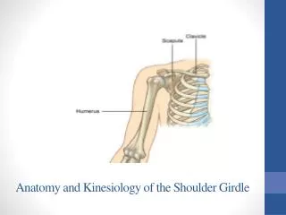
Bones of the Elbow The lower end of the humerus flairs out as epicondyles. These provide a mechanical advantage to the forearm muscle groups that attach at these sites. Lateral Medial Medial lateral extensors flexors extensors capitulum trochlea Anterior view Posterior view
FRONT OF FOREARM • MUSCLES • 5 superficial muscles : all cross elbow joint • 3 deep muscles : do not cross elbow joint • Flexors = more bulky - antigravity muscles & used for grip • ARTERIES • Superficial radial; deep ulnar • Anterior interosseous a. (branch of ulnar) supplies deepest structures on front of forearm • NERVES • Median nerve (with its ant. Interosseous branch) is main nerve of front of forearm • Radial nerve (with its post. Interosseous branch) is main nerve of back of forearm
SUPERFICIAL MUSCLES : FRONT OF FOREARM • Pronator Teres (PT) • Flexor Carpi Radialis (FCR) • Palmaris Longus (PL) • Flexor Carpi Ulnaris (FCU) • Flexor Digitorum Superficialis (FDS)
Superficial Muscles of the Anterior Forearm • 5 superficial muscles • From the common flexor tendon arising from the medial condyle of the humerus • Cross the elbow but have no function at that joint
Surface Anatomy - Anterior Forearm lateral medial Thumb = pronator teres 5th digit (tucked under) = flexor digitorum superficialis 2nd digit = flexor carpi radialis 3rd digit = palmaris longus 4th digit = flexor carpi ulnaris
SUPERFICIAL MUSCLES : FRONT OF FOREARM • ADDITIONAL ORIGINS • Deep (ulnar) head of PT • Medial margin of coronoid process of ulna • Ulnar head of FCU • Medial margin of olecranon • FDS (ulnar & radial heads) • Ulnar head • Sublime tubercle on medial border of cornoid process, • Radial head • Anterior border of radius
INSERTIONS • PT • middle 1/3 lateral surface shaft of radius • FCR • base 2nd & 3rd mertacarpal bones • PL • flexor retinaculum; • FCU • pisiform bone • FDS • forms 4 tendons for medial 4 fingers; opposite bases of proximal phalanges each tendon is perforated by tendon of FDP; split tendon inserts into 2 sides of shaft of middle phalanx
FRONT OF FOREARM • NERVE SUPPLY • FCU : ulnar nerve • PT, FCR, PL, FDS : median nerve • ACTION • PT : main pronator of forearm & hand • FCR, PL, FCU : chief flexors of wrist • FCR : abducts hand • FCU : fixes pisiform bone during contraction of hpyothenar muscles; abducts little finger
Deep Muscles of the Anterior Forearm • 3 Deep Muscles Thumb Fingers Wrist • Arise from the ulna (pronator quadratus, flexor digitorum profundus) and radius (flexor pollicis longus)
FRONT OF FOREARM : FDP • Origin • Upper anterior & medial surfaces of shaft of ulna • Medial surfaces of olecranon & coronoid processes of ulna • Insertion • Palmar surface of base of distal phalanges • Innervation • Medial 1/2 - ulnar nerve • Lateral 1/2 - anterior interosseous nerve • Action • Flexion of distal phalanges
FRONT OF FOREARM : FPL • Origin • Upper 3/4 of anterior surface of shaft of radius • Adjoining part of anterior surface of • interosseous membrane • Insertion • Palmar surface of base of distal phalanx of thumb • Innervation • Anterior interosseous nerve • Action :Flexes distal phalanx of the thumb
PRONATOR QUADRATUS • Origin • lower 1/4 of anterior surface of shaft of ulna • Insertion • lower 1/4 of anterior surface of radius • Innervation • Anterior interosseous nerve • Action • Superficial fibers pronate forearm • Deep fibers bind lower ends of radius & ulna PRONATOR QUADRATUS
Median Nerve • All forearm muscles are innervated by the MEDIAN nerve EXCEPT: 1 ½ muscles • flexor carpi ulnaris • ulnar side of the flexor digitorum profundus Plus: All thenar mm except adductor pollicis
Radial & Ulnar Arteries medial lateral Radial artery Ulnar artery Common interosseous • Anterior • Posterior Dorsal and palmer carpal branches Dorsal and palmer carpal branches Deep (superficial) palmar arches superficial (deep)palmar arches
EXTENSOR COMPARTMENT : BACK OF FOREARM 12 muscles 1. Anconeus 2. Brachioradialis (BR) 3. Extensor carpi radialis longus (ECRL) 4. Extensor carpi radialis brevis (ECRB) 5. Extensor digitorum (ED) 6. Extensor digiti minimi (EDM) 7. Extensor carpi ulnaris (ECU) 8. Supinator (Sup) 9. Abductor pollicis longus (APL) 10. Extensor pollicis brevis (EPB) 11. Extensor pollicis longus (EPL) 12 Extensor indicis (EI)
Superficial Posterior Muscles • Brachioradialis • Extensor carpi radialis longus • Extensor carpi radialis brevis • Extensor digitorum • Extensor carpi ulnaris • Anconeus
Superficial muscles Extensor side Extensor retinaculum Brachioradialis (flexor) Anconeus Ext. carpi rad. longus Ext. carpi rad. brevis Ext. carpi ulnaris Ext. digit. comm. Ext. digiti minimi
Deep Posterior Muscles of the Forearm • Supinator • Abductor pollicis longus • Extensor pollicis brevis and longus • Extensor indicus
Deep muscles Extensor side Abductor pollicis longus Ext. indicis Ext. pollicis brevis Extensor pollicis longus
Origin • All 7 superficial muscles cross elbow joint • Arise from tip of lateral epicondyle of humerus • 5 deep muscles arise from 2 bones of forearm & interosseous membrane
EXTENSOR RETINACULUM Definition : Deep fascia of back of wrist thickened for holding extensor tendons Attachments : Laterally: to the lower part of the anterior border of radius Medially: (1) the styloid process of ulna (2) triquetral (3) pisiform bones
EXTENSOR COMPARTMENT : NERVE SUPPLY Anconeus BR ECRL Radial nerve ECRB ED EDM ECU Sup APL EPB EPL EI Posterior interosseous nerve
superficial radial n. radial a. abductor pollicis longus tendon extensor pollicis brevis tendon extensor pollicis longus tendon 1st dorsal interosseus m. Anatomical snuff box: ABD, EPB, EPL