Chapter 11 DNA and Protein Synthesis
380 likes | 585 Vues
Chapter 11 DNA and Protein Synthesis. Mrs. Svencer. 11.1 Genes are made of DNA. Frederick and Griffith Transformation –bacteria with mice Strain A – pneumonia, fatal Strain B – harmless Heated strain A – harmless Mix heated strain A and strain B - death Harmless bacteria - “transformed”

Chapter 11 DNA and Protein Synthesis
E N D
Presentation Transcript
Chapter 11DNA and Protein Synthesis Mrs. Svencer
11.1 Genes are made of DNA • Frederick and Griffith • Transformation –bacteria with mice • Strain A – pneumonia, fatal • Strain B – harmless • Heated strain A – harmless • Mix heated strain A and strain B - death • Harmless bacteria - “transformed” • Descendents were too - trait was passed on • Heritable change
Figure 11-1Griffith showed that although a deadly strain of bacteria could be made harmless by heating it, some factor in that strain is still able to change other harmless bacteria into deadly ones. He called this the "transforming factor."
Oswald Avery – focused on protein and DNA • Heat strain A + strain B + protein-destroying enzymes • Offspring still transformed • Protein not accountable for transformation • Heat strain A + strain B + DNA-destroying enzymes • Colonies did not transform • Therefore, DNA = genetic material
Hershey and Chase – used viruses • Virus = nucleic acid in a protein coat • Bacteriophage = virus that infects bacteria • Batch 1 • Labeled protein coats with radioactive sulfur • Radioactivity detected outside of the cells • Batch 2 • Labeled DNA with radioactive phosphorus • Radioactivity inside cells • Therefore, phage’s DNA entered bacteria • Therefore, DNA is the hereditary material
Figure 11-4Hershey and Chase offered further evidence that DNA, not proteins, is the genetic material. Only the DNA of the old generation of viruses is incorporated into the new generation.
11.2 Nucleic Acids store information • DNA (deoxyribonucleic acid) • Stores genetic information • Built from nucleotides – 3 parts • 1. Sugar – ring shape, “deoxyribose” • 2. Phosphate group • 3. Nitrogenous base – single or double ring of C and N atoms
Figure 11-5A nucleotide has three components: a sugar, a phosphate group, and a nitrogenous base.
4 bases in DNA • Pyrimidines: single rings • Thymine (T) • Cytosine (C) • Purines: double rings • Adenine (A) • Guanine (G)
Figure 11-6DNA contains four different nitrogenous bases. Thymine and cytosine have single-ring structures. Adenine and guanine have double-ring
DNA Strands • Covalent bonds connect sugar to phosphate between nucleotides • Sugar-phosphate “backbone” • Nucleotides arrange in different • 1. Numbers • 2. Sequences • (combinations are unlimited)
Rosalind Franklin and Maurice Wilkins • X-ray crystallography • DNA = helix shape • Watson and Crick • Made model of double helix using Franklin’s pictures • Twisted ladder
Complementary Base Pairs • Purine + Pyrimidine • A-T (2 hydrogen bonds) • G-C (3 hydrogen bonds)
11.3 DNA Replication – mechanism of inheritance (DNA – copying) • Template Mechanism • 2 strands of double helix separate at origins of replication • Copying goes outward from origin in both directions, making a “bubble” • Each strand is a “template” for a new, complementary strand • Bases line up according to base-pairing rules
Figure 11-9During DNA replication, the two strands of the original parent DNA molecule, shown in blue, each serve as a template for making a new strand, shown in yellow. Replication results in two daughter DNA molecules, each consisting of one original strand and one new strand.
Figure 11-10DNA replication begins at origins of replication and proceeds in both directions, producing "bubbles." Eventually, all the bubbles merge, resulting in two separate daughter DNA molecules.
DNA polymerase (enzyme) links nucleotides together using covalent bonds • DNA opens further as nucleotides added • Eukaryotic DNA – many origins - speedy • New strands = daughter strands • Fast and accurate • 1 error / billion nucleotides
Review: DNA Replication occurs before a cell divides during the S phase of interphase of the cell cycle
11.4 Genes Provide Information for Making Proteins • Beadle and Tatum – orange bread mold • Mutant strains unable to grow on nutrient medium • Lacked single enzyme needed to produce a needed molecule • Each mutant defective in a single gene • “one gene-one enzyme” hypothesis – one gene produces one specific enzyme • NOW, “one gene – one polypeptide”
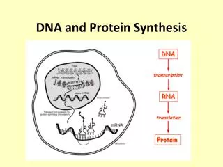
Gene to Protein • 1. Transcription - DNA to RNA • Transcribed message (RNA) leaves nucleus to cytoplasm • 2. Translation – RNA to amino acid • Codon = on RNA - 3 bases that code for amino acid
Table of Codons • RNA • Figure 11-13 • 61/64 code for amino acids – some amino acids coded for by more than 1 codon • Ex: UUU UUC – both code for phenylalanine • 3/64 – stop codons – end of each gene sequence
Figure 11-13Each codon stands for a particular amino acid. (The table uses abbreviations for the amino acids, such as Ser for serine.) The codon AUG not only stands for methionine (Met), but also functions as a signal to "start" translating an RNA transcript. There are also three "stop" codons that do not code for amino acids, but signal the end of eachgenetic message.
11.5 Two steps from Gene to Protein Transcription: DNA to RNA • Messenger RNA (mRNA) transcribed from DNA template – only 1 strand of DNA • RNA nucleotides pair to complementary bases – AUCG • RNA polymerase - RNA nucleotides together • RNA splicing – introns are removed and exons are joined together • Therefore, mRNA has a continuous coding sequence • mRNA leaves nucleus
Figure 11-12Information flows from gene to polypeptide. First, a sequence of nucleotides in DNA (a gene) is transcribed into RNA in the cell's nucleus. Then the RNA travels to the cytoplasm where it is translated into the specific amino acid sequence of a polypeptide.
Figure 11-15In eukaryotes, the RNA transcript is edited before it leaves the nucleus. Introns are removed and the exons are spliced together before the "final draft" transcript moves into the cytoplasm where it gets translated.
Intron = noncoding region of mRNA • Exon = coding region of mRNA that is “expressed” or translated
Translation: RNA to protein • 1. Start codon: AUG –translation begins • 2. Amino acids added one-by-one to a chain of amino acids • A. tRNA (transfer RNA) translates codons of mRNA to amino acids • 1. tRNA molecule binds to appropriate amino acid
2. tRNA recognizes, using base-pairing rules, codons in mRNA by its own complementary anticodon • Anticodon = 3 bases at one end of tRNA • 3. Other end of tRNA = where amino acid attaches • 4. An enzyme links tRNA to its amino acid, using ATP
Figure 11-16During translation, tRNAs transport and match amino acids to their appropriate codons on the mRNA transcript. One end of the tRNA attaches to an amino acid. At the other end, a triplet of bases called the anticodon matches to the complementary mRNA codon.
B. occurs at ribosome • 2 subunits – made of protein and rRNA (ribosomal RNA) • Small subunit – binding site for mRNA • Large subunit – 2 binding sites for tRNA • 1. “P” site (polypeptide) – holds tRNA carrying the growing polypeptide chain • 2. “A” site (amino acid) – holds tRNA carrying next amino acid to be added to the chain • 2 subunits hold mRNA and tRNA close together • C. ribosome connects new amino acid to the growing polypeptide chain
Figure 11-17Ribosomes bring mRNA and tRNAs together during translation. Each ribosome has an attachment site for an mRNA transcript, and two sites for tRNAs.
3. Reaches stop codon • UAA, UAG, UGA • No amino acid at “A” site • Translation stops • 4. Completed polypeptide set free from tRNA by hydrolysis
11.6 Mutations • Mutation – any change in the nucleotide sequence of DNA • Can be • Large regions of chromosomes • Single nucleotide pairs
Base substitutions • Replacement of 1 base or nucleotide with another • Sometimes no effect – “silent mutation” • Sometimes large effect • Why? • Several amino acids have more than 1 codon
More disastrous mutations • Insertion • Putting in an additional nucleotide • Deletion • Taking away 1 nucleotide • Both insertion and deletion alter the triplet groupings • Now they code for new amino acids
Cause of mutations • Error during DNA replication • Error during crossing over in meiosis • Mutagens – physical / chemical agents that cause mutations • Ex: high-energy radiation, x-rays, UV light • Usually harmful, sometimes helpful • Ex: dark color in female tiger swallowtails
Mutations may be passed to offspring, if they occur in gametes • Ultimate source of genetic diversity