Virological Methods
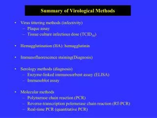
Virological Methods. Viral infectivity : ~ Plaque forming unit (PFU), ~ Tissue culture 50% infective dose (TCID 50 ) Serology ( antibody response to the virus) Molecular methods ~ Polymerase chain reaction (PCR) Virus specific methods ~ Hemagglutination

Virological Methods
E N D
Presentation Transcript
Virological Methods • Viral infectivity: • ~ Plaque forming unit (PFU), • ~ Tissue culture 50% infective dose (TCID50) • Serology ( antibody response to the virus) • Molecular methods • ~ Polymerase chain reaction (PCR) • Virus specific methods • ~ Hemagglutination • ~ MAGi test for HIV (human immunodeficiency virus) • Electron Microscope
EM • Mechanism: accelerate electrons to high energy, then focuses them using a magnet. Because virus particles are stained with osmium (heavy metal), electron will not pass through them, and it allows the visualization of the virus particles. • Advantages: count the number of the virus particles in a sample regardless of infectious or not. • Cryo EM: for enveloped viruses • X-ray crystallograph: visualization of viral proteins, computer methods is usually used to reconstruct the image based on the known structure
EM pictures and the reconstructed X-ray crystallograph images • Morphology of virus (EM) • Symmetry of nucleocapsid structure (X-ray) • Not all the viruses (need high concentrations of purified virus) • Attachment-receptor interaction (X-ray structure of glycoproteins)
Plaque assay Mechanism: based on the cytopathic effects (CPE) of viruses ~ use of a confluent monolayers of cells which are susceptible to the virus ~ induction of visible cytopathic effects (cell killing,or cell damage ~ semisolid overlay (agar) which prevents virus diffusion from one infected cell to the other nearby cells ~ small round plaques (clear areas) visualized by staining cells with a vital dye like neutral red or crystal vioilet
Calculation of PFU • Serial 10-fold dilution of virus sample are tested(duplicates) • e.g 10-1-10-10 (depending on the virus one is working with • and the information about the aliquot to estimate the range • of virus dilution) • The dilution of virus which gives 20-100 plaques per petri dish • is used for calculation of PFU • e.g 10-5 dilution gives 20 plaques, and 0.5 ml of virus • dilution is added to the cell monolayers, then the PFU of • the virus stock will be 20 x 105 , equals 4 x 10⁶PFU/ml 0.5 ml
Virus particles vs. plaque forming particles EM: virus particles regardless of infectivity Plaque assay: Virus particles with infectivity
TCID50 • Mechanism: similar to plaque assay, based on the CPE of virus and measures the infectivity of virus particles • For viruses which do not form distinct plaque on infected cells • Use a statistic method for quantification • Similar procedures as plaque assay, but liquid medium is used for the incubation, no agar is added to the medium, no staining is needed, observe CPE under light microscope • Minimal 5 duplicate wells for each dilution. Usually 10 well/dilution
Calculation of TCID50 • Use of Reed & Muench method (if 50 ul of virus dilution is added to eachwell)
Hemagglutination assay • Mechanism: Viruses have hemagglutinin as glycoprotein to • agglutinate red blood cells of certain species animals (enveloped viruses, such as influenza virus) • Measures the hemagglutination units, not virus particles, not • infectivity ( minimal virus required to agglutinate red blood cells in a well is called one unit)
Procedures • Do a serial two-fold dilution of virus sample • Mix with certain fixed amount of red blood cells • Incubate at room temperature for 30 minutes • Observe with naked eye, virus containing wells appear as lattice, No virus wells appear as “button”
MAGI assay for HIV • Mechanism: use of indicator cells which contain virus specific inducible reporter gene • MAGI cell system for HIV • Susceptible to HIV • Engineered to contain HIV LTR sequence and E.coli-galactosidase gene (LacZ) • HIV infection > tat protein > LTR promoter > LacZ > stain with x-gal > blue cells • Others use green fluoresence protein (GFP) as reporter gene • Virus specific proteases: reporter gene fusion protein, virus > protease > cleave fusion protein > blue (LacZ) or green (GFP) • Reverse transcriptase assay for retroviruses and hepatitis B virus
Serological Methods • Mechanism: use antibody to detect viral proteins or use viral • proteins to detect host immune responses • ~ Virus neutralization: defining virus serotype (e.g. • Polio virus type 1,2 and 3, VSV NJ vs. IN). • ~ Immunofluorescence staining (IFA): detecting viral • proteins with labeled antibody (direct) or detecting • antibody with a labeled secondary antibody (indirect) • e.g.,labeled anti-mouse antibody is a secondary, initial • mouse anti-viral antibody is the primary antibody • ~ ELISA and immunoprecipitation methods
Virus neutralization • Mechanism: Antibody is added to a virus preparation, allow to • react for one hour, then measure the infectivity of the preparation using plaque assay or TCID50. • VN titer: calculated by comparing the PFU/ml or TCID50 of virus preparation with and without reacting with the antibody • Two methods: • ~10-fold dilution of virus preparation with constant amount of antibody • ~ 10-fold dilution of antibody with constant amount of virus
IFA • Direct and indirect • Simple, diagnostic • Specificity depend on the antibody used (monoclonal antibody better) • Background staining
Immunoprecipitation • Detect viral proteins • Radioactive label (35-Met) virus infected cells • Cell lysates react with viral specific antibody • Protein A remove antigen-antibody complex • SDS-PAGE: protein gel electrophoresis • Very sensitive, little amount of viral proteins
Immunoblot method • Detect viral proteins • Not label with radioactive materials • Whole cell lysates subjected to SDS-PAGE • Transfer to solid membrane support (nitrocellulose membrane) • Probed with specific antibody conjugated with indicator molecule (horse radish peroxidase) • Observe with a chromgen
Enzyme-linked immunosorbent assay (ELISA) • Similar to IFA • Direct (measure antigen) • Indirect (measure antibody) • A solid matrix, plastic plate • Same detection methods as immunoblot (indicator molecule such as horse radish peroxidase and a chromgen) • Highly specific and sensitive • Diagnostic (HIV)
Nucleic acid detection • Polymerase chain reaction (PCR): DNA viruses • Reverse transcriptase(RT)-PCR: RNA viruses • Real-time PCR: quantitative • Primers, template, DNA polymerase, dNTP, PCR reaction buffer • Reverse transcriptase for RT-PCR • Real-time PCR: probes
PCR Amplification profile Amplication vs amount of input DNA Denature (95oC) Annealing (45oC-70oC) Extension (72oC)
Real-time PCR • Taqman assay • Quench dye reduce the emission of reporter dye • Release of reporter dye is measured
Pulse-chase experiment • Radioactive material labeling of virus infected cells (35S-Met) • Pulse: labeling with radioactive materials • Chase: different time periods after labeling, cell lysates prepared for SDS-PAGE • Autoradiograph to detect labeled proteins • Kinetics, dynamic of viral protein processing 35S-Met Pulse for 5 minutes wash chase 5 min 10 min 20 min 40 min 1 hour Fresh medium