Download
1 / 134
1.35k likes | 2.32k Vues
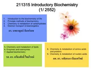
211315 Introductory Biochemistry (1/ 2552). Introduction to the biochemistry of life Principle methods of biochemistry Chemistry & metabolism of carbohydrates Electron transport & bioenergetics. ดร. นพกาญจน์ จันทร์เดช. 5. Chemistry and metabolism of lipids 6. Enzymes and coenzymes

E N D
211315 Introductory Biochemistry (1/ 2552) • Introduction to the biochemistry of life • Principle methods of biochemistry • Chemistry & metabolism of carbohydrates • Electron transport & bioenergetics ดร. นพกาญจน์ จันทร์เดช 5. Chemistry and metabolism of lipids 6. Enzymes and coenzymes 7. Applied biochemistry 8. Chemistry & metabolism of amino acids and proteins 9. Chemistry & metabolism of nucleic acids รศ. ดร. เกรียงศักดิ์ ไชยโรจน์ ผศ. ดร. หทัยชนก เนียมทรัพย์
หนังสืออ่านประกอบ 1) ชีวเคมีทั่วไป 2) Biochemistry: the chemistry of life (International Edition). David T. Plummer 3) Harper’s Biochemistry (25th-edition). Robert K. Murray, Daryl K. Granner, Peter A. Mayes, and Victor W. Rodwell 4) General, Organic and Biochemistry (4th-edition). Denniston, K.J., Topping, J.J., and Caret, R.L.
OBJECTIVES • เข้าใจหลักการและวิธีการตรวจวัดขั้นพื้นฐานทางชีวเคมี • ชนิด และลักษณะสำคัญของ carbohydrates • Metabolism ของ carbohydrate ชนิดต่างๆ ในสิ่งมีชีวิตชนิดต่างๆ • กลไกการถ่ายทอดอิเลคตรอนในสิ่งมีชีวิต และพลังงานในสิ่งมีชีวิต
1) Introduction to Biochemistry of Life • What is biochemistry? • Categories of biochemistry • Chemical materials of life = Biomolecules; carbohydrate (sugar • and polysaccharides, protein (amino acid and enzymes), lipid, • nucleic acid • Reaction of biomolecules = Metabolism (anabolism and catabolism) • 3) Metabolic regulation
protein, carbohydrate, lipid, enzyme, nucleic acid glucose, amino acid C H O N [S] Macromolecules Molecules atom Metabolism (metabolism) Catabolism NADH or NADPH Micro molecules Lactic acid Pyruvic acid CO2 , NH3 Urae Macro molecules Carbohydrate Polysaccharides sugar Protein Lipid Oxidative phosphorelation Reducing equivalence Energy O2 H2O Anabolism
Metabolic Pathway (metabolic pathway) • Catabolic pathway: • การหายใจของเซลล์; สร้างพลังงาน (ATP และ NADPH) • Aerobic respiration ได้แก่ Electron transport chain Oxidative phosphorylation • Anaerobic respiration ได้แก่ Cori cycle Lactic acid fermentation Ethanol fermentation • Glycogenolysis; สลายแป้ง (แป้งในสัตว์)/ไกลโคเจน กลูโคส • - Glycolysis; กลูโคส ไพรูเวต (pyruvate) และ ATP • - Entner-Doudoroff Pathway;alternative-glycolysis ในแบคทีเรีย • - Pentose phosphate pathway (hexose monophosphate shunt); สร้าง NADPH จากกลูโคส • Protein catabolism; protein amino acid
Anabolic pathway; เป็นกระบวนการสร้างโมเลกุลของสารที่ซับซ้อน จากสารประกอบตั้งต้นอย่างง่าย: - Glycogenesis: การสังเคราะห์ไกลโคเจน - Gluconeogenesis: การสังเคราะห์กลูโคส - Secondary metabolism; กระบวนการสร้างสารที่ไม่สำคัญต่อการเจริญเติบโต แต่มีความสำคัญต่อการดำรงชีวิต - การสังเคราะห์แสง (Photosynthesis) ปฏิกิริยาที่ขึ้นกับแสง (light reaction) ปฏิกิริยาที่ไม่ขึ้นกับแสง (dark reaction)
DNA Nucleus Prokaryotic cell Eukaryotic cell ในเซลล์แบคทีเรียมี Mitochondria !! Anatomy of the bacterial cell
ลักษณะสำคัญของ Prokaryotic cell (page2-3) • 1 ไม่มีเยื่อหุ้มนิวเคลียส • 2. เยื่อหุ้มเซลล์มีหน้าที่หลายอย่าง • ลักษณะสำคัญของ Eukaryotic cell • มีเยื่อหุ้มนิวเคลียส • มีcytoskeleton; microtubules & microfilaments • มี exocytosis & endocytosis • มีการแบ่งเซลล์แบบ mitosis & miosis • ลักษณะพื้นฐานที่เหมือนกัน • มีเยื่อหุ้มเซลล์ 2. มีDNA เป็นสารพันธุกรรม 3. มีเอนไซม์ควบคุม • มีRNA เป็นตัวถ่ายทอดคำสั่งจาก DNA 5. สังเคราะห์โปรตีนที่ไรโบโซม • 6. ใช้ ATP เป็นแหล่งพลังงานเบื้องต้น
Protoplasm cytoplasm nucleus Cell wall (ไม่มีในเซลล์สัตว์; เกิด plasmolysis) Cell membrane (semipermeable membrane) Organelles Endoplasmic Recticulum (ER) RER (สังเคราะห์โปรตีน) & SER (สลายไกลโคเจน) Lysosome (มีเฉพาะในเซลล์สัตว์) Ribosome (สังเคราะห์โปรตีน) Mitochondria** Plastid (ไม่พบในรา แบคทีเรียcyanobacteria) leucoplast (สะสมอาหาร) chromoplast (รงควัตถุ) chloroplast (รงควัตถุสีเขียว) grana stroma Vacuole sap vaculoe/ food vacuole/ pinocytotic vacuole/ contractile vacuole/ nucleolar vacuole/ gas vacuole centriole L
2) Principle Methods of Biochemistry 2.1 sample preparation: (p-8) Breaking up cells/ tissue Homogenization Centrifugation 2.2 separation methods: (p-9-14) Chromatography paper and thin layer chromatography column chromatography Ion exchange chromatography gel filtration HPLC gas chromatography Electrophoresis 2.3 analytical methods: (p-15)
centrifugation นำของเหลวภายในcell ไปวิเคราะห์ ** ชนิด, ปริมาณ, ทำให้บริสุทธิ์ Cell lysis • Osmotic pressure • Freeze & thaw • Sonication แยกน้ำเลี้ยงกับ cell ออกจากกันได้ 2.1 Sample preparation
Centrifugation: “centrifugal force” เป็นแรงเหวี่ยงที่ทำให้อนุภาคหนีออกจากจุดศูนย์กลาง ระดับของcentrifugal force ขึ้นกับขนาดของอนุภาค macromolecule จะเคลื่อนออกจากจุดศูนย์กลางได้เร็วกว่าmicromolecule หน่วยของความเร็วในการ centrifuge: times gravity (×g) หรือ centrifugation rotor speed; revolution per minute (rpm) g = (1.118 × 10⁻⁵) R S2 g = times gravity R = รัศมีของหัวเหวี่ยง (cm) S = speed of the centrifuge in rpm (revolution per minute) Example, centrifugation of a sample at 5,000 rpm in a microcentrifuge that has a rotor with a radius of 7 cm will deliver a centrifugal force of 1,957 × g
Types of rotor 1. Fixed angle rotors (14-40゚, usually 30゚) 2. Swing-out rotors (used for pelleting small quantities of materials) 3. Vertical rotors 4. Continuous flow rotors (for cell harvesting)
Cell debris (เศษเซลล์) Organelles Ribosome Fractional centrifugation 10 min, 600xg 10 min, 10,000xg 180 min, 100,000xg
Separation & Purification • a) Chromatography • หลักการ สารต่างชนิดกันมีความสามารถในการละลายใน mobile phase • และละลายใน Stationary phase ต่างกัน • Paper chromatography • Stationary phase = น้ำในกระดาษกรอง • Mobile phase = solvent or buffer (ที่มีขั้วน้อยกว่าน้ำ) • การแยกของสารผสมที่มีสี สามารถมองเห็นได้ด้วยตาเปล่า • กรณีสารผสม เป็นสารไม่มีสี • -Thin layer material + fluorescent indicator ; UV-detectable • -Compound Color reaction • Ex. unsaturated compound :: Iodine vapour • amino acid :: Ninhydrin reagent • reducing sugar:: Dinitrosalicylic acid, Benedict’s solution
Solvent front Filter paper Thin layer plate Solvent front Solvent 1 2 3 1 2 3 Sample 1 2 3 Fig. Schematic of TLC Fig. Separation by TLC Rf = ระยะที่สารเคลื่อนที่ (cm) ระยะที่ตัวทำละลาย(ตัวชะ)เคลื่อนที่ (cm) Rf value depends on the nature of the thin layer material, and the composition of the solvent
- Column chromatography ตัวชะ (eluent) ตัวกลาง (stationary phase) หลักการ eluent จะเป็นตัวพาสารผสมออกจาก columnทางด้านล่างด้วยความเร็วต่างๆกัน ประสิทธิภาพของ column chromatography ขึ้นอยู่กับชนิดของสารตัวอย่าง, การเลือกใช้ตัวกลาง และขนาดของ column
Ion-exchange chromatography • - สารตัวกลางเป็นสารที่มีประจุ เช่น carboxymethylcelluloseซึ่งเป็น • celluloseที่มี carboxymethyl group (-CH2COO-) หรือ polystylene • ที่มี aminomethyl group(-CH2NH3+) • - สารตัวกลาง = resin หรือexchanger หรือ matrix • - ประจุในตัวกลางทำหน้าที่ยึดสารที่มีประจุตรงข้ามไว้ • anion exchange chromatography ; สารตัวกลางมีประจุบวก (แลกเปลี่ยนประจุลบ) • เช่น diethylaminoethyl-;DEAE -O-C2H5-NH-C2H5 • C2H5 • cation exchange chromatography ; สารตัวกลางมีประจุลบ (แลกเปลี่ยนประจุบวก) • เช่น carboxymethyl- ; CM -O-CH2-COO-
OCH2COO- CM-Cellulose OCH2COO- กำหนดให้ตัวชะ คือ KCl Cellulose Cation exchanger OCH2COO- OCH2COO- OCH2COO- A- KCl Samples ; OCH2COO- OCH2COO- B+ B+ K+ OCH2COO- Cl- A- K+ (ความเข้มข้นต่ำ)**
pH 7.0 ตัวอย่าง: การชะโปรตีนผสมออกจาก column 1. pH gradients 2. Ionic strength gradient ** pH gradients Decrease pH of buffer for anion-exchange chromatography(ประจุลบ กลายเป็นกลาง) Increase pH of buffer for cation-exchange chromatography(ประจุบวกกลายเป็นกลาง) pH 6.0 pH 5.0 Phenylalanine Protein#1 Protein#2 Protein#3 Protein Detection: Abs 280 nm Anion-exchange chromatography (target protein = negative)
ตัวอย่าง: การชะโปรตีนผสมออกจาก column 1. pH gradients 2. Ionic strength gradient ** Ionic strength gradients KCl (0.5M) KCl (1.0M) KCl (low conc. 10 mM) Prot1 (+++) Prot2 (++) Prot3 (+) Prot4 (-) Prot1 (+++) Prot2 (++) Prot1 (+++) Prot3 (+) Prot4 (-) Prot2 (++) Cation-exchange chromatography (target protein = negative)
A280 nm Prot2 (++) [KCl] M 2.0 Prot1 (+++) Prot3 (+) & prot4 (-) 1.5 1.0 0.5 Fraction no.
- Gel filtration - แยกสารต่างๆได้ตามขนาดโมเลกุลของสาร - ตัวกลางคือ polysaccharideเช่น sephadex, sepharose, agaroseเป็นต้น - ตัวกลางมีคุณสมบัติยอมให้โมเลกุลขนาดเล็กลอดผ่านเข้าไปได้ - เมื่อชะสารต่างๆออกจาก column สารที่มีโมเลกุลเล็กจะเคลื่อนที่ออกจาก columnได้ช้า - พิจารณาขนาดโมเลกุลของสารตัวอย่าง, ชนิดของตัวกลาง และความยาวของ column
Gas chromatography (GC) • -ใช้แยกสารพวก volatile non-polar substances โดยเปลี่ยนสารผสมให้เป็นไอที่อุณหภูมิหนึ่ง • แล้วผ่านไอไปสู่ column • - stationary phase = gas or liquid (gas-liquid chromatography) • ถ้า stationary phase เป็นพวก non-polar จะเสถียรที่ช่วงอุณหภูมิกว้าง • - mobile phase = gas เช่น Nitrogen, Helium, Hydrogen • การวิเคราะห์ • sample injection • สารผสมจะถูกให้ความร้อนจนกลายเป็นไอแล้วถูกพาเข้าไปใน column • ตรวจวัดโดย detector และแสดงออกมาในรูปของ chromatogram
High Performance Liquid Chromatography (HPLC) • - Stationary phase: ของเหลวที่เคลือบอยู่บนสารยึดซึ่งเป็นของแข็งบรรจุใน columnขนาดเล็ก • Sampleจะถูกฉีดผ่าน injectorและอาศัยแรงดันจากปั๊มช่วยนำตัวทำละลาย (mobile phase) • ออกจาก reservoir พาsample ผ่าน column • -ระยะเวลาที่sample ผ่านเข้าออก columnเรียกว่า retention time • ในสภาวะเดียวกัน สารชนิดเดียวกันจะมีretention time เท่ากัน • สามารถหาชนิดของสารตัวอย่างได้จากการเปรียบเทียบ retention timeกับสารมาตรฐาน • สามารถหาปริมาณของสารตัวอย่างได้จากการคำนวณพื้นที่ใต้peak เทียบกับสารมาตรฐานที่ทราบชนิด • และปริมาณ
ตัวอย่าง Standard glucose 10 mM Standard fructose 10 mM Standard sucrose 10 mM
การหาปริมาณกลูโคสในสารตัวอย่างการหาปริมาณกลูโคสในสารตัวอย่าง Glucose 100 mM Detection signal Glucose 10 mM Glucose 1 mM Retention time พื้นที่ใต้กราฟ glucose (unit 2) 1 10 100 ความเข้มข้น glucose (mM)
b) Electrophoresis เป็นการแยกสารโดยอาศัยกระแสไฟฟ้า หลักการ อนุภาคที่มีประจุไฟฟ้าจะเคลื่อนที่ไปในสนามไฟฟ้า อนุภาคที่มีประจุบวกจะ เคลื่อนที่ทวนทิศทางของสนาม ส่วนอนุภาคประจุลบจะเคลื่อนที่ตามทิศทางของสนาม macromoleculeที่มีหมู่ -NH3+, -COO-, หรือ -PO32- จะมีประจุบนโมเลกุล ทิศทางกระแสไฟฟ้า - ตัวกลาง = กระดาษกรอง(with buffer) - เหมาะสำหรับวิเคราะห์สารโมเลกุลเล็ก -หลังจากผ่านกระแสไฟฟ้าแล้ว นำกระดาษไปผึ่งให้แห้ง - ถ้าสารที่แยกได้ไม่มีสีอาจตรวจดูได้โดยการย้อมสี หรือทำปฏิกิริยาให้เกิดสี เช่น ถ้าเป็นกรดอะมิโน อาจพ่นด้วยนินไฮดริน แล้วทำให้ร้อน จะเกิดสีน้ำเงิน
Polyacrylamide gel electrophoresis (PAGE) ตัวกลาง = polymer ของสาร acrylamideและ N,N’-methylene-bis-acrylamide -เจลมีลักษณะพิเศษคือ เส้นpolymerมีลักษณะเป็นตาข่าย ดังนั้นจึงสามารถกั้นโมเลกุลขนาดใหญ่ให้ เคลื่อนที่ช้าลงได้ ดังนั้นอัตราการเคลื่อนที่ของสารจึงถูกกำหนดด้วยประจุ และขนาดโมเลกุล -ในกรณีที่มี sodium dodesylsulfate (SDS) ซึ่งเป็นผงซักฟอกอยู่ในระบบจะทำให้สารละลาย โปรตีนที่ต้องการแยกเสียสภาพ และมีประจุต่อหน่วยมวลใกล้เคียงกัน (all negative) ดังนั้น การแยกโปรตีนด้วยวิธี SDS-PAGE จะแยกสารออกจากกันโดยอาศัยขนาดโมเลกุลเท่านั้น - สามารถใช้ SDS-PAGE หาน้ำหนักโมเลกุลของโปรตีนได้
Polyacrylamide gel electrophoresis Tris buffer pH 9.0
c) Spectroscopy คือการวัดปริมาณรังสีแม่เหล็กไฟฟ้า ที่สารหนึ่งสามารถดูดซับเอาไว้ได้เมื่อมี รังสีฉายผ่านสารนั้น รังสีแม่เหล็กไฟฟ้า อาจมีความยาวคลื่น (wavelength) ได้ ต่างๆกัน เรียกว่า spectrum - สารต่างชนิดกันมีความสามารถในการดูดซับรังสีที่ความยาวคลื่นต่างกัน - สารชนิดเดียวกัน ความเข้มข้นหรือปริมาณต่างกัน ดูดกลืนแสงที่ความยาวคลื่นเดียวกันได้ไม่เท่ากัน - หน่วยความยาวคลื่น คือ นาโนเมตร (nm) - UV light: 185 – 400 nm Visible light: 400-800 nm
เครื่องมือและหลักการที่ใช้ใน spectroscopy spectrophotometerหรือ spectrometerคือเครื่องมือที่ใช้วัดการดูดกลืนรังสี แม่เหล็กไฟฟ้าของสาร ประกอบด้วย แหล่งของรังสี และส่วนที่ใช้เลือก ความยาวคลื่น (monochrometer) รังสีจากแหล่งจะผ่าน monochrometerไปยังสารละลาย ซึ่งจะดูดเอาไว้ส่วนหนึ่ง รังสีส่วนที่ทะลุออกมาสามารถวัดได้ด้วย detector
กฎของเบียร์-แลมเบิร์ตกฎของเบียร์-แลมเบิร์ต • ความเข้มของรังสีที่มาตกกระทบ (I0) จะลดลงเป็น I เมื่อผ่านสารละลายเป็นระยะทางdx • “ ความเข้มของรังสีที่ถูกดูดเอาไว้(dI)จะแปรเป็นสัดส่วนโดยตรงกับความเข้มข้น (c)และระยะทางที่รังสีนั้นวิ่งผ่านสารละลาย (dx)” • ʃdI = ʃk’cdx ; k’ = ค่าคงที่ • I • ---- ถ้า conc. มาก อัตราส่วนระหว่าง dI/I มาก ---- • ---- ถ้า dx มาก อัตราส่วนระหว่าง dI/I มาก ---- I d dx I0 0 I I0
ดังนั้น ถ้าให้รังสีความเข้มข้น I0วิ่งผ่านสารละลายเป็นระยะทาง dx จะได้ log (Io/I) = kcdx log(Io/I) =optical density (OD)หรือ absorbance (A)ของสารละลาย k(เท่ากับ k’.ln10ซึ่งเรียกว่า extinction coefficientจะมีค่าคงที่เมื่อความยาวคลื่นคงที่ และเป็นค่าจำเพาะสำหรับสารนั้นๆ ** ถ้าความเข้มข้น (c) เป็นโมลาร์ และ dx มีหน่วยเป็นซม. จะเรียกค่า k ว่า molar extinction coefficient, εมีประโยชน์ในการใช้หาความเข้มข้นของสารจาก OD เมื่อทราบεของสารนั้น
A = ε c l A = absorption of solution ε=molar extinction coefficient (M-1 cm-1) l = path length of cuvette (cm) C = concentration (M) cuvette ตัวอย่าง สารละลาย tryptophan มีค่าการดูดกลืนแสง0.55ที่ 280 nmใน cuvette ที่มีความหนา 0.5 cm , εของ tryptophanเท่ากับ 5600 M-1 cm-1 สารละลายนี้มีความเข้มข้นเท่าไหร่ C = A / (ε d) = 0.55/ (5600 . 0.5) = 1.96 x 10⁻⁴ M
- สารชีวโมเลกุลที่เป็น aromatic หรือมี conjugated double bond สามารถดูดกลืนรังสีในแถบ ultraviolet (185-350 nm) ได้ - carotene, hemoglobin, chlorophyll สามารถดูดกลืนแสงในช่วง visible light - nucleotide ซึ่งเป็นส่วนประกอบของ nucleic acid สามารถดูดกลืนรังสี Ultraviolet ได้ดีที่สุดที่ 260 nm (λmax = 260 nm)
PHOTOSYNTHESIS 6CO2 + 6H2O C6H12O6 + 6O2 Plants are consumed by animals Animals release CO2 + H2O ENERGY METABOLISM 6O2+ C6H12O6 6CO2 + H2O + energy and BIOSYNTHESIS 3) Chemistry of Carbohydrates -C:H:O = 1:2:1 -General formula; (CH2O)n, n= 3-7 “monosaccharides” -Polysaccharides; starch, cellulose, glycogen
Dihydroxyaldehyde (aldose) Dihydroxyacetone (ketose) Type of carbohydrates Monosaccharides -General formula; (CH2O)n, n= 3-7 -monosaccharides can be named on the basis of the functional group (carbonyl group) -monosaccharides contain many “Hydroxyl group (-OH)” polyhydroxylaldehydes/ polyhydroxylketones
Monosaccharides: nomenclature • Functional group: aldose ketose • Carbon atom: triose (C-3), tetrose (C-4), pentose (C-5), hexose (C-6)** • Unique name: e.g., glucose (aldo-hexose) fructose (keto-hexose) galactose (…) glyceraldehyde (…)
D- and L-configuration of monosaccharides -the prefixes D and L found in the complete name of monosaccharide are used to identify one of two possible isomeric forms called “stereoisomers” “ stereoisomers …same molecular formula, same bonding, but different in arrangement ” Single asymmetric carbon or Chiral carbon
น้ำตาลที่มี chiral center มากกว่า 1 ตำแหน่ง จะพิจารณาD, Lจาก asymmetric Cที่อยู่ไกลจาก aldehyde or keto group มากที่สุด Most naturally occurring sugars are D isomers. D & L sugars = mirror Images, same name
Enantiomers: Stereoisomers ที่สมมาตรกันเหมือนส่องกระจก เช่น D-glucose กับ L-glucose Diastereoisomers: Stereoisomers ที่ไม่มีคุณสมบัติเป็น Enantiomers Epimers: Diastereoisomers ที่มีตำแหน่ง chiralC ที่สมมาตรกันเพียง 1 ตำแหน่ง
Enantiomers HO H Epimers
การพิจารณา D-, L- configuration ไม่เกี่ยวข้องกับ การหมุนระนาบแสง (optical rotation) Levorotatory sugar (S- หรือ (-)- sugar): หมุนไปทางซ้าย ทวนเข็มนาฬิกา Dextrorotatory sugar (R- หรือ (+)-sugar): หมุนไปทางขวา ตามเข็มนาฬิกา สามารถพบกลูโคสทั้งแบบที่เป็น D(+) glucose และ D(-) glucose L(+) glucose และ L(-) glucose
จำนวน isomers เท่ากับ 2n ; n คือจำนวน chiral carbon