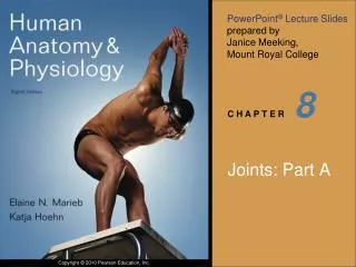
8
8 . Joints: Part A. Joints (Articulations). Articulation—site where two or more bones meet Functions of joints: Give skeleton mobility Hold skeleton together. Classification of Joints . There are two ways to classify joints: Functional and Structural

8
E N D
Presentation Transcript
8 Joints: Part A
Joints (Articulations) • Articulation—site where two or more bones meet • Functions of joints: • Give skeleton mobility • Hold skeleton together
Classification of Joints • There are two ways to classify joints: Functional and Structural • Functional Classification of joints is based on amount of movement allowed by the joint • Synarthroses—immovable • Amphiarthroses—slightly movable • Diarthroses—freely movable
Classification of Joints • Structural Classification of Joints - Based on material binding bones together and whether or not a joint cavity is present (Fibrous, Cartilagenous, Synovial) • Three types of fibrous joints: • Sutures-occur only between skull bones; jagged articulations w/ short fibers btwn. • Syndesmoses- bones connected by ligaments, cords/bands of CT • Gomphoses –a peg in socket joint; the only example is a tooth in its bony alveolar socket
(a) Suture Joint held together with very short, interconnecting fibers, and bone edges interlock. Found only in the skull. Suture line Dense fibrous connective tissue Figure 8.1a
(b) Syndesmosis Joint held together by a ligament. Fibrous tissue can vary in length, but is longer than in sutures. Fibula Tibia Ligament Figure 8.1b
(c) Gomphosis “Peg in socket” fibrous joint. Periodontal ligament holds tooth in socket. Socket of alveolar process Root of tooth Periodontal ligament Figure 8.1c
Classification of Joints • Cartilagenous Joints: • Two types of cartilagenous joints • Synchondroses- a bar of hyaline cartilage unites bone; mostly all are synarthrotic; (ex. epiphyseal plates in children) • Symphyses – articular surfaces of bones covered with hyaline which is fused to an intervening pad of fibrocartilage (ex. intervertebral discs)
(a) Synchondroses Bones united by hyaline cartilage Sternum (manubrium) Epiphyseal plate (temporary hyaline cartilage joint) Joint between first rib and sternum (immovable) Figure 8.2a
(b) Symphyses Bones united by fibrocartilage Body of vertebra Fibrocartilaginous intervertebral disc Hyaline cartilage Pubic symphysis Figure 8.2b
Synovial Joints General Structures of Synovial Joints • Articular cartilage: pad of hyaline cartilage on ends of long bones • Joint (synovial) cavity: small potential space; contains synovial fluid • A double-layered joint capsule : an outer fibrous capsule (dense irregular CT) and an inner synovial membrane (loose CT)
Synovial Joints General Structures of Synovial Joints • Synovial fluid- a viscous fluid occupying joint cavity; lubricates and nourishes articular cartilage • May have wedges of fibrocartilage separating the articular surfaces called: menisci • Fibrous (bursae) lined with: synovial membrane contains synovial fluid and acts as friction reducing ball bearings
Ligament Joint cavity (contains synovial fluid) Articular (hyaline) cartilage Fibrous capsule Articular capsule Synovial membrane Periosteum Figure 8.3
Subacromial bursa Humerus resting Cavity in bursa containing synovial fluid Humerus moving Figure 8.4b
Types of Synovial Joints • Plane-Nonaxial joints, short gliding movements • Hinge -Uniaxial joints, flexion and extension only • Pivot – Uniaxial joints, rotation • Condyloid - Biaxial joints, permit all angular movements • Saddle – Biaxial, more movement than condyloid • Ball and socket - Multiaxial joints; the most freely moving synovial joints
f Nonaxial Uniaxial Biaxial Multiaxial c b Plane joint (intercarpal joint) a a e d Figure 8.7a
f Nonaxial Uniaxial Biaxial Multiaxial c b Hinge joint (elbow joint) b a e d Figure 8.7b
f Nonaxial Uniaxial Biaxial Multiaxial c b c Pivot joint (proximal radioulnar joint) a e d Figure 8.7c
f Nonaxial Uniaxial Biaxial Multiaxial c b d Condyloid joint (metacarpophalangeal joint) a e d Figure 8.7d
f Nonaxial Uniaxial Biaxial Multiaxial c b e Saddle joint (carpometacarpal joint of thumb) a e d Figure 8.7e
f Nonaxial Uniaxial Biaxial Multiaxial c b f Ball-and-socket joint (shoulder joint) a e d Figure 8.7f
Common Joint Injuries • Sprains - Ligaments are stretched or torn; slow to repair themselves • Cartilage tears - Due to compression and shear stress, fragments may cause joint to lock or bind; Cartilage rarely repairs itself • Dislocations – Bones are forced out of alignment
Torn meniscus Figure 8.14
Inflammatory and Degenerative Conditions • Bursitis - Inflammation of a bursa, usually caused by a blow or friction • Tendonitis - Inflammation of tendon sheaths typically caused by overuse
Arthritis • Symptoms; pain, stiffness, and swelling of a joint • Acute forms: caused by bacteria, treated with antibiotics • Chronic forms: osteoarthritis, rheumatoid arthritis, and gouty arthritis
Gliding Movements • One flat bone surface glides or slips over another similar surface • Examples: • Intercarpal joints • Intertarsal joints • Between articular processes of vertebrae
Gliding (a) Gliding movements at the wrist Figure 8.5a
Angular Movements • Movements that occur along the sagittal plane: • Flexion—decreases the angle of the joint • Extension— increases the angle of the joint • Hyperextension—excessive extension beyond normal range of motion
Hyperextension Extension Flexion (b) Angular movements: flexion, extension, and hyperextension of the neck Figure 8.5b
Extension Hyperextension Flexion (c) Angular movements: flexion, extension, andhyperextension of the vertebral column Figure 8.5c
Flexion Extension Flexion Extension (d) Angular movements: flexion and extension at theshoulder and knee Figure 8.5d
Angular Movements • Movements that occur along the frontal plane: • Abduction—movement away from the midline • Adduction—movement toward the midline • Circumduction—flexion + abduction + extension + adduction of a limb so as to describe a cone in space
Abduction Circumduction Adduction (e) Angular movements: abduction, adduction, andcircumduction of the upper limb at the shoulder Figure 8.5e
Rotation • The turning of a bone around its own long axis • Examples: • Between C1 and C2 vertebrae • Rotation of humerus and femur
Rotation Lateral rotation Medial rotation (f) Rotation of the head, neck, and lower limb Figure 8.5f
Special Movements • Movements of radius around ulna: • Supination - lateral rotation of hands • Pronation - medial rotation of hands
Pronation (radius rotates over ulna) Supination (radius and ulna are parallel) (a) Pronation (P) and supination (S) Figure 8.6a
Special Movements • Movements of the foot: • Dorsiflexion - upward movement • Plantar flexion - downward movement
Dorsiflexion Dorsiflexion Plantar flexion Plantar flexion (b) Dorsiflexion and plantar flexion Figure 8.6b
Special Movements • Movements of the foot: • Inversion - turn sole medially • Eversion - turn sole laterally
Inversion Eversion (c) Inversion and eversion Figure 8.6c
Special Movements • Movements in a transverse plane: • Protraction- anterior movement • Retraction-posterior movement
Protraction of mandible Retraction of mandible (d) Protraction and retraction Figure 8.6d
Special Movements • Elevation - lifting a body part superiorly • Depression - moving a body part inferiorly
Depression of mandible Elevation of mandible (e) Elevation and depression Figure 8.6e
Special Movements • Opposition of the thumb -Movement in the saddle joint so that the thumb touches the tips of the other fingers
Opposition (f) Opposition Figure 8.6f