Protein Structure: From Pauling and Corey to X-Ray Crystallography
This article provides an overview of protein structure, including the importance of proteins in cellular function, the process of protein synthesis, and the use of X-ray crystallography in determining protein structures. It also highlights the contributions of Linus Pauling and Robert Corey in elucidating protein structures.

Protein Structure: From Pauling and Corey to X-Ray Crystallography
E N D
Presentation Transcript
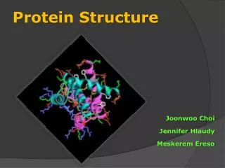
Protein Structure (in a nutshell) Guy Ziv December 26th, 2006 Myoglobin (1958)
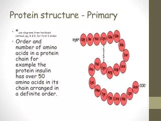
Proteins • From the Greek “proteios” meaning “of first importance” • The basic building blocks of almost all life • Constitutes the majority of the cell, and perform nearly all enzymatic activities • Composed of 20 naturally occurring amino-acids varying moiety called “side chain”
Protein Synthesis In-vivo • Transcription: DNA messenger RNA (mRNA) • Translation: mRNA Linear chain of a.a.(Ribosome) • Folding:Linear chain Structure Peptide bond Protein chains have direction N-terminal → C-terminal
X-Rays crystallography – the tool of structural biology • Why X-ray?Wavelength of visible light: ~500 nmBond lengths in proteins: ~0.15 nmTypical X-ray wavelength: ~0.15 nm • X-ray are (weakly) scattered by electrons • Diffraction from a single molecule is weakso use a crystal: • Multiple copies of the molecule increases diffraction • Crystalline structure imposes constraints on diffraction pattern
q q q Diffraction occurs at particular angles • Diffraction spots are the result of constructive interference from multiple scatterers satisfying Bragg’s Law: λ = 2 d sinθ θ
Bragg planes intersect the unit cell in particular “indices” 0,0 k h h=1, k=1 h=4, k=-2
Each Spot Represents a Unique Set of Bragg Planes Points in k-space (Fourier Space) h=2, k=1, l=3 h=10, k=3, l=8 detector λ = 2 d sinθ q1 q2 q3
Modern X-Ray Crystallography Need good crystals for better resolution, which is difficult in proteins (need right conditions) and sometimes nearly impossible (e.g. membranal proteins) Early 1950’s High resolution details are faint – requires good experimental apparatus Recorded intensity give only the magnitudebut not the phase of the complex “form factor” Error in density map lead to un-realistic atom assignment, requiring iterative refinement process
Historical perspective to Pauling and Corey paper series • X-ray crystallography, invented in the beginning of the 20’th century, has been used to solve structures of some amino-acids, synthetic polymers (poly-glu) and small organic molecules • Some fibrous materials such as wool and α-keratin are sufficiently crystalline to give diffraction patterns • Evidence suggested that these proteins’ structure involve mainly translation and rotation
Pauling and Corey Robert Corey (1897-1971) Linus Pauling (1901-1994)
Pauling and Corey papers series –PNAS April 1951 • Pauling, L., Corey, R.B. and Branson H. R. The Structure of Proteins: Two Hydrogen-Bonded Helical Configurations of the Polypeptide Chain. PNAS, 37, 205-211, (1951). • Pauling, L. & Corey, R. B. Atomic Coordinates and Structure Factors for Two Helical Configurations of Polypeptide Chains. PNAS, 37, 235-240, (1951). • Pauling, L. & Corey, R. B. The Structure of Synthetic Polypeptides. PNAS, 37, 241-250, (1951). • Pauling, L. & Corey, R. B. The Pleated Sheet, A New Layer Configuration of Polypeptide Chains. PNAS, 37, 251-256, (1951). • Pauling, L. & Corey, R. B. The Structure of Feather Rachis Keratin. PNAS, 37, 256-261, (1951). • Pauling, L. & Corey, R. B. The Structure of Hair, Muscle, and Related Proteins. PNAS, 37, 261-271, (1951). • Pauling, L. & Corey, R. B. The Structure of Fibrous Proteins of the Collagen-Gelatin Group. PNAS, 37, 272-281, (1951). • Pauling, L. & Corey, R. B. The Polypeptide-Chain Configuration in Hemoglobin and Other Globular Proteins. PNAS, 37, 282-285, (1951).
Pauling and Corey papers series –PNAS April 1951 • Pauling, L., Corey, R.B. and Branson H. R. The Structure of Proteins: Two Hydrogen-Bonded Helical Configurations of the Polypeptide Chain. PNAS, 37, 205-211, (1951). • Pauling, L. & Corey, R. B. Atomic Coordinates and Structure Factors for Two Helical Configurations of Polypeptide Chains. PNAS, 37, 235-240, (1951). • Pauling, L. & Corey, R. B. The Structure of Synthetic Polypeptides. PNAS, 37, 241-250, (1951). • Pauling, L. & Corey, R. B. The Pleated Sheet, A New Layer Configuration of Polypeptide Chains. PNAS, 37, 251-256, (1951). • Pauling, L. & Corey, R. B. The Structure of Feather Rachis Keratin. PNAS, 37, 256-261, (1951). • Pauling, L. & Corey, R. B. The Structure of Hair, Muscle, and Related Proteins. PNAS, 37, 261-271, (1951). • Pauling, L. & Corey, R. B. The Structure of Fibrous Proteins of the Collagen-Gelatin Group. PNAS, 37, 272-281, (1951). • Pauling, L. & Corey, R. B. The Polypeptide-Chain Configuration in Hemoglobin and Other Globular Proteins. PNAS, 37, 282-285, (1951).
Linus Carl PaulingThe Nobel Prize in Chemistry 1954 "for his research into the nature of the chemical bond and its application to the elucidation of the structure of complex substances"
Determinants of helical structure superposition Resonant partial double bond character of peptide bond induces planar arrangement of atoms Distances and angles Between atoms All hydrogen bonds should be satisfied, i.e. distance N-O of about 2.7Å and anglebetween C = O and H – N less then ~30°
Building a model – similar to building with LEGO blocks • Start assembling monomers (amino-acids) with fixed translation and rotation • Look for configurations which have no steric hindrance (i.e. clashes) • Calculate N-H…O=C distances andangles (3-d trigonometry..)
The α-helix – one of the two common structural elements in proteins • Completes one turn every 3.7 residues • Rises ~5.4 Å with each turn • Has hydrogen bonds between the C=O of residue i and the N-H of residue i+4 • Is right-handed N C i O i+4
Alpha-helices appear a lot in trans-membranal proteins membrane E.g. Lactose permease (LacY)
Why did Pauling and Corey succeed where others failed? • Understanding the importance of hydrogen bonds • Taking into account the planar peptide bond • Better knowledge of covalent bond lengths and angles • MOST IMPORTANTLY – they were NOT crystallographers, and did not consider only models with integer number of residues per turn!
Proof came 7 years later… John Cowdery Kendrew The Nobel Prize in Chemistry 1962 Kendrew, J. C., Bodo, G., Dintzis, H. M. Parrish, R. G., Wyckoff, H., and Phillips, D. C. A Three-Dimensional Model of the Myoglobin Molecule Obtained by X-ray Analysis. Nature, 181, 662 (1958).
Hierarchy of Protein Structure Linear chain made of 20 possible amino acids Alpha-helices, beta-sheets, turns Motifs, domains Oligomers, complexes
The PDB contains over 40,000 structures (as of December 2006) NMR - Nuclear magnetic resonance Allows structure determination based on distance and angular constraints in solution
Proteins’ Structure is Dynamic • Fluctuations exists in all proteins • Conformational changes ↔ Function Adenylate kinase An enzyme that catalyzes the production of ATP from ADP
Protein Folding – still an open question • 1954 Christian B. Anfinsen proved that the protein structure is determined by it’s sequenceProtein Denatured (unfolded) Protein • 1969 “Levinthal paradox” – For a 100 a.a. sequence there are 9100 possible configurations. If sampled randomly every nanosecond, it will take longer then the age of the universe to fold a single protein + Urea Dilution RNase enzyme
Protein folding – research continues • Late 1980’s - Wolynes et al. present the “Energy Landscape” or “Folding Funnel” model for protein folding • 2006 – There is still no precise understanding how proteins fold fast (up to µsec!), reliably and accurately to their native structure Entropy Energy Native (folded) state