Download
1 / 35
380 likes | 456 Vues
Explore the intricate details of the male reproductive system, from internal development to hormonal regulation. Learn about spermatogenesis, semen production, and the role of testosterone. Dive into the mechanisms and effects of testosterone activity and hormonal regulation, shedding light on male fertility.

E N D
Reproductive PhysiologyThe Male Reproductive System Dr. Khalid Alregaiey
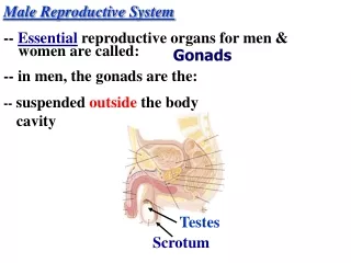
Sexual Differentiation: Internal Embryonic Development • Bipotential tissues: genes & hormones direct differentiation • Gonad testis or ovary • Wolffian duct Vas deferens, • Mullerian duct oviduct
Regulation of Reproduction: General Pathways • Hypothalamus: pulse generator • Gonadotropin releasing H (GnRH) • Anterior Pituitary • Lutenizing H (LH) • Follicle stimulating H (FSH) • Ovary: • Estrogen, progesterone, Inhibin • Testis: testosterone
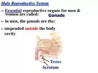
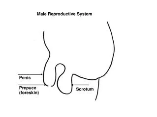
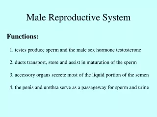
Male Reproductive Anatomy and Physiology • Testis • Epididymis • Vas deferens • Seminal vesicle • Prostate • Bulbourethral • Ejaculatory duct • Urethra • Penis
Sertoli Cells • Form blood-testes barrier: • Prevents autoimmune destruction of sperm. • Produce FAS ligand which binds to the FAS receptor on surface to T lymphocytes, triggering apoptosis of T lymphocytes. • Prevents immune attack. • Secrete inhibin. • Phagocytize residual bodies: • May transmit information molecules from germ cells to Sertoli cells. • Secrete androgen-binding protein (ABP): • Binds to testosterone and concentrates testosterone in the tubules.
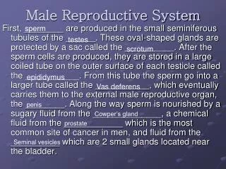
Spermatogenesis • Seminiferous tubules • Contain spermatogonia • Stem cells involved in spermatogenesis • Contain Sertoli (sustentacular) cells • Sustain and promote development of sperm
Spermiogenesis • Maturation of spermatozoa. • Phagocytosis of cytoplasm by the Sertoli cells. • Cytoplasm is eliminated. Insert fig. 20.18
Regulation of Spermatogenesis • GnRH LH Leydig cells testosterone growth and division of germ cells • GnRH FSH Sertoli cells spermatoctye maturation • Inhibin feedback – FSH, testosterone – short & long loops • Estrogen • Growth hormone
Maturation of Sperm in Epididymis • Sperms in the early portion of epididymis are nonmotile • After 18-24 h they develop capability of motility • Most of sperms are stored in epididymis • After ejaculation they become motile • Activity of a sperm is greatly enhanced in neutral to slightly alkaline medium
Seminal Vesicles • S Vs produce fructose, citric acid and other nutrients as well as prostaglandins and fibrinogen Prostate • Slightly alkaline milky fluid that help in neutralizing other seminal fluids as well as the vaginal fluids • Prostates fluids also contain clotting protein and profibrinolysin
Semen • Milky white, sticky mixture of sperm and accessory gland secretions (65% of semen is from seminal vesicle, 25% prostate) • Provides a transport medium and nutrients (fructose), protects and activates sperm, and facilitates their movement • pH is 7.5 • Prostaglandins in semen: • Decrease the viscosity of mucus in the cervix • Stimulate reverse peristalsis in the uterus • Facilitate the movement of sperm through the female reproductive tract
Semen • Clotting factors coagulate semen immediately after ejaculation, then fibrinolysin liquefies the sticky mass during the next 15-30 minutes • After ejaculation, sperms can live 24-48 h • Freshly ejaculated semen undergoes a process called capacitation: 1. inhibitory factors are washed out by uterine and fallopian fluids, 2. the sperm swims away from cholesterol vesicles, 3. the mebrane of the sperms becomes more permeable to Ca++
Semen • Only 2-5 ml of semen are ejaculated, but it contains 35-200 million sperm/ml (<20 million infertile) • When the majority of the sperm are morphologically abnormal or nonmotile then person is likely to be infertile
Hormonal Regulation of Testicular Function • The hypothalamus releases gonadotropin-releasing hormone (GnRH) • GnRH stimulates the anterior pituitary to secrete FSH and LH • FSH causes Sertoli cells to release androgen-binding protein (ABP) • LH stimulates interstitial (Leydig) cells to release testosterone • ABP binding of testosterone enhances spermatogenesis
Hormonal Regulation of Testicular Function • Feedback inhibition on the hypothalamus and pituitary results from: • Rising levels of testosterone • Increased inhibin
Mechanism and Effects of Testosterone Activity • Testosterone is synthesized from cholesterol • It binds to testosterone –binding globulin (TeBG), ABP, serum albumin, or to corticosterone-binding globulin (CBG) • Once it diffuses to cells it either binds to androgen receptor or converted to DHT which then binds to the androgen receptor • Testosterone targets all accessory organs, its deficiency causes these organs to atrophy • It causes testes descent during the last 2-3 months of gestation.
Testosterone Functions: • Testosterone targets all male reproductive organs and accessory glands, its deficiency causes these organs to atrophy • Causes the appearance of pubic, axillary, and facial hair • Enhances growth of the chest and deepening of the voice • Skin thickens and becomes oily • Bones grow and increase in density and calcium retention. It is also responsible for the male pelvis shape (narrow, long, funnel-like shape).
Testosterone functions (continued) • It increases basal metabolic rate • Increases red blood cells • It also causes hair growth (pubic, axillary) and libido in females. • Spermatogenesis and erection.
Male Sexual Act • Erection is initiated by sexual stimuli including: • Touch and mechanical stimulation of the glans penis and other parts • Erotic sights, sounds, and smells • Erection can be induced or inhibited solely by emotional or higher mental activity • Enlargement and stiffening of the penis from engorgement of erectile tissue with blood
During sexual arousal, a parasympathetic reflex promotes the release of nitric oxide, VIP, and Acetylcholine. • Nitric oxide relaxes the penis arteries and causes erectile tissue to fill with blood • Expansion of the corpora cavernosa: • Compresses their drainage veins • Retards blood outflow and maintains engorgement
When the sexual stimulus becomes extremely intense, spinal cord begins to send sympathetic impulses to initiate emission • Filling of the internal urethra with semen elicits signals that promotes ejaculation • After orgasm, the excitement disappears within 1-2 minutes (resolution)