Download
1 / 17
170 likes | 380 Vues
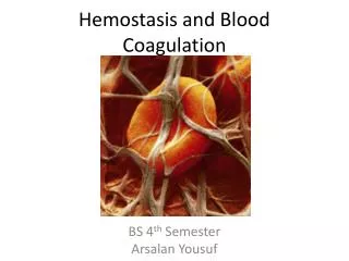
Influence of Blood Flow on the Coagulation Cascade. Nina Marianne Andersen, Mads Peter Sørensen, Emil Sokoler Department of Mathematics (MAT), Techn. Univ. of Denmark Steen Ingwersen and Ole Hvilsted Olsen Biomodelling and Haemostasis Biochemistry, Novo Nordisk, Denmark. Content:

E N D
Influence of Blood Flow on the Coagulation Cascade Nina Marianne Andersen, Mads Peter Sørensen, Emil Sokoler Department of Mathematics (MAT), Techn. Univ. of Denmark Steen Ingwersen and Ole Hvilsted Olsen Biomodelling and Haemostasis Biochemistry, Novo Nordisk, Denmark Content: Introduction, blood coagulation. Perfusion experiment, cartoon model and reaction schemes in a fully stirred model. Cartoon model and reaction schemes for a simplified model with diffusion and flow. Platelet activation. Numerical results and relation to other models. Summary and future work plan.
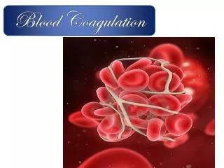
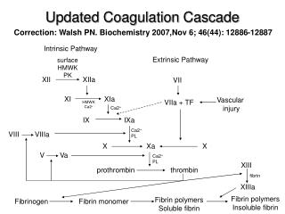
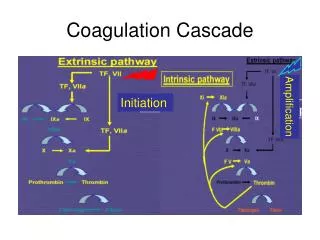
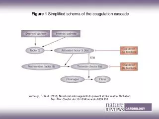
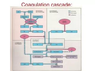
Cartoon of the blood coagulation pathway. Ref: http://www.ambion.com/tools/pathway/pathway.php?pathway=Blood%20Coagulation%20Cascade
Perfusion experiments and modelling Perfusion chamber Active thombocytes (Ta) binds to the collagen coated lid. vWF. Top glass lid coated with collagen Factor X in the fluid phase X Thrombocytes (platelets), red and white blood cells. Factor VIIa in the fluid phase VIIa Reconstructed blood. Content: Thrombocytes (T), Erythrocytes. [T] = 14 nM (70,000 platelets / μ litre blood)
Cartoon model of the perfusion experiment UnactivatedPlatelet ActivatedPlatelet IIa IIa II IIa Va:Xa VIIa X Xa V Va Activated Platelet
Reaction schemes, one example. Factor II (prothrombin): II Factor IIa (thrombin): IIa Prothrombinase complex: Xa_Va_Ta A total of 17 equations. Reaction rates: Ref: P.M. Didriksen, Modelling hemostasis - a biosimulation project, internal report, Dept. 252 Biomodelling, Novo Nordisk
Numerical results. Initial conditions: FVIIa = 50 nM FX = 170 nM T = 14 nM sTa = 0.1*14 nM FII = 0.3 nM IIa T VIIa Ta
Reaction diffusion model with convection Reaction scheme for T, Ta and IIa. Corresponding model equations in the space Ω. Poiseuille’s flow
Boundary conditions and parameters Boundary condition x=0 Boundary condition x=l: Outflow boundary conditions. Top and bottom boundary condition: No flow crossing. Ref.: M. Anand, K. Rajagopal, K.R. Rajagopal. A Model Incorporating some of the Mechanical and Biochemical Factors Underlying Clot Formation and Dissolution in Flowing Blood. Journal of Theoretical Medicine, 5: 183-218, 2003.
Numerical results. Time = 0.6 sec. T IIa T-IIa Ta
Numerical results. Time = 5 sec. T IIa T-IIa Ta
Numerical results. Time = 10 sec. T IIa T-IIa Ta
Future work: Boundary attachment of Ta Reaction schemes on Corresponding model equations on.
One simple example including diffusion and flow Reaction scheme in the bulk: Reaction scheme on parts of the boundary: Conservation of binding sites at the boundary: Boundary binding sites:
Including pro-coagulant and anti-coagulant thrombin Ref.: V.I. Zarnitsina et al, Dynamics of spatially nonuniform patterning in the model of blood coagulation, Chaos 11(1), pp57-70, 2001. E.A. Ermakova et al, Blood coagulation and propagation of autowaves in flow, Pathophysiology og Haemostasis and Thrombosis, 34, pp135-142, 2005.
Summary and future work Modelling of perfusion experiment for blood coagulation. Reduced PDE model including blood flow and diffusion. Modelling of attachment of activated thrombocytes on collagen coated boundary. Full PDE model. Model of in vivo blood coagulation.