Download
1 / 76
1.01k likes | 2.63k Vues
Assessment of myocardial viability. Viability Dysfunctional myocardium subtended by diseased coronary arteries Limited or absent scarring Potential for functional recovery Prospective definition Reversible ischemic contractile dysfunction Myocardial stunning Myocardial hibernation.

E N D
Viability • Dysfunctional myocardium subtended by diseased coronary arteries • Limited or absent scarring • Potential for functional recovery • Prospective definition • Reversible ischemic contractile dysfunction • Myocardial stunning • Myocardial hibernation
Myocardial stunning • Heyndrickx et al 1978 • Prolonged and fully reversible contractile dysfunction of the ischemic heart that persists after reperfusion • Transient period of ischemia f/b reperfusion-depressed function at rest,preserved perfusion • Affected area responsive to inotropes • Time course not altered by use of inotropes-spontaneously resolve within a week • Duration of stunning depends on the duration and severity of ischemia and the adequacy of arterial flow
Clinical relevance • Exercise induced ischemia • Acute cor.syndrome • Angioplasty-balloon inflation • Cardiopulmonary bypass
Mechanism • Calcium hypothesis-decrease responsiveness to calcium • Oxyradical hypothesis-ROS during reperfusion impairs calcium handling
Myocardial hibernation • Diamond et al 1978 • Persistent LV contractile dysfunction when myocardial perfusion is chronically reduced but sufficient to maintain viability of tissue • Depressed function and perfusion at rest • Progressively reversible after revascularisation • Time to restoration- • Months to one year • Depend on duration and severity of flow reduction&ultrastructural changes
Mechanisms • Smart heart hypothesis • Myocardial function &metabolism reduced to match a reduction in blood flow • Repetitive stunning hypothesis • Repetitive episodes of ischemia reperfusion from supply demand mismatch leading to sustained depression of contractile function
Cellular mechanisms • Apoptosis prominent during transition to hibernation-30%cell loss • Compensatory regional myocyte hypertrophy-to maintain normal wall thickness • Increase in interstitial connective tissue,myolysis,increased glycogen deposition,minimitochondria • Cell survival programme-downregulation of glycogen synthasekinase,increase anti apoptotic proteins • Downregulation of beta adrenergic adenylylcyclase
Clinical assessment • Heart failure and active angina • Directly angiography • Viability study may have a role in planning revascularisation strategy once coronary anatomy known • Heart failure and no angina • Class 1 recommendation for assessment of viability in pts with CAD and LVD • Class 2a rec.for assessment of co-presence of CAD
STEMI • Class2a rec. for viability assessment 4 to 10 days after STEMI in hemodynamically&electrically stable pts.to define potential effect of revascularisation
Strong association b/w myocardial viability and improved survival after revascularization in pts with chronic CAD and LV dysfunction. • Allmann KC et al;JACC 2002 • Pts with viability-revascularization a/w 79% reduction in mortality (16% vs. 3.2%) as compared to conserv.Rx • Pts without viability-no significant difference in revasc. Vs medical therapy (7.7% vs. 6.2%)
In patients with ischemic cardiomyopathy 55% had viable myocardium by PET&27% had improved LVEF after revascularisation • Auerbach MA et al;circulation 1999
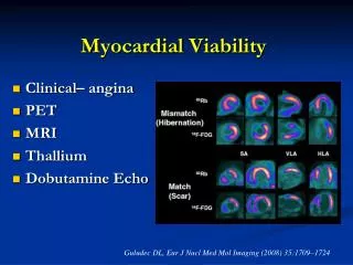
Techniques for assessment of viability • Myocardial glucose utilisation-PET FDG • Cell membrane integrity-SPECT Thallium • Intact mitochondria-SPECT Tc • Contractile reserve-dob.echo and dob.MRI • Scar tissue-DEMRI,MSCT
Echocardiographic assessment • LV end-diastolic wall thicknesses • Less than 6 mm-less likely to be viable
Dobutamine stress echo • Low-dose dobutamine (5–10 μg/kg/min) • Increase contractility in viable myocardium • High-dose dobutamine(upto 40 μg/kg/min) • Biphasic response –initial improvement F/B worsening –underperfused but viable tissue-most specific sign of improvement after revasc. • Uniphasic response-sustained improvement-myocardial damage with subsequent reperfusion-less predictive of improvement after revasc. • Deterioration of wall motion without initial improvement-severe ischemia • No change in wall motion-scar • Sensitivity(84%),specificity(81%)for recovery of function
Strengths- • Higher specificity • Viability&ischemia assessed • MR can be detected • Good spatial resolution • Widely available • Lower cost • Predictive of clinical outcomes
Limitations • Poor window in 30% • Lower sensitivity • Viable regions with absent flow reserve will not show thickening • Reliance on visual assessment
Myocardial contrast echo • Gas-filled microbubbles <7 μ as contrast agents • Produces myocardial opacification and facilitates identification of LV borders • Burst of high-intensity ultrasound-destroy microbubbles within the myocardium-replenishment observed over the next 10 to 15 cardiac cycles • Viable if homogeneous contrast intensity • Absence of contrast enhancement-nonviable myocardium
Sensitivity 89% and specificity of 51% • Higher sensitivity,lower specificity compared to DSE
Strengths • Microcirculatory integrity • Extent of viability • Precise deleination • Viability assessed in total occlusion • Limitation • Poor window • Scant clinical data
Myocardial strain rate imaging • Determination of velocities in two segments of myocardium separated by a distance • Strain rate is rate of change of velocity b/w these points • Increased sensitivity from 73% to 83% compared to visual assessment
Echo based techniques • Strengths • Safety, portability, low cost,widespread availability of equipment • Absence of radiation hazard • Limitations • Operator dependent • Spatial resolution is relatively low • Diagnostic accuracy reduced in poor acoustic window
TMT • Exercise induced ST elevation in infarct related area was associated with residual tissue viability • Margonoto et al ;JACC 1995 • Sensitivity&specificity of reciprocal ST-segment depression a/w exercise-induced ST-segment elevation in prior Q wave infarct for detecting residual viability-84%&100% • Nakano A et al;JACC 1999
SPECT-Th-201 • K+ analogue utilizes active cellular transport system-relies on intact cell • Uptake depends on viability ®ional perfusion • Redistribution-gradual accumulation of tracer in hypoperfusedareas,rapid washout from normally perfused areas • Segments with tracer uptake >60%-viable • Subendocardial scar tissue may be labelled as viable-lower specificity
Rest redistribution protocols- • Defects in initial images that improve in 4 hour image-viable myocardium • Additional 24 hr image if fixed defects in 4 hr image • S/L NTG prior to injection • Less sensitive-86%,specificity 47%
Rest redistribution thallium-defect in inferoseptal wall that redistributes
Stress redistribution protocol • Pharmac.or exercise stress • Th inj. &imaging f/b 4 hr image • Myocardium not perfused with rest or stress-scar • Defect on stress and improves on rest-ischemic &viable • 24 hr image for late redistribution
Stress redistribution reinjection protocols • Reinjection of Th201 and image repeated 24hr later • Viable myocadium-uptake of tracer on reinjection in segments with no uptake on stress • Scar –defect that persists • Sensitivity 90% specificity 54%
Tc99m labelled agents • Rely on sarcolemmal integrity and mitochondrial function • Short half life,higherdoses,better image • Redisribution less-less helpful in assessing viability • Tc NOET-similar redistribution kinetics to Thallium • Viability criterion is>50% tracer uptake in dysfunctional segments.
Ant Apex Stress Inf Rest Septum Lateral Stress Apex Rest Lat Inferior Anterior Sep Ant Stress Lat Sep Inf Rest Apex Base Reversible Ischeamia, defect appears at stress and disappears during rest
Ant Apex Stress Inf Rest Septum Lateral Stress Apex Rest Lat Inferior Anterior Sep Ant Stress Lat Sep Inf Rest Apex Base Fixed Scar, defect is seen in both stress and rest
Strengths • High sensitvity • Quantitative objective criteria • LVEF • FDG with special collimator • Predictive of outcomes
Limitations • Reduced spatial resolution &sensitivity compared to PET • Attenuation artefacts • Cannot differentiate endocardial viability • Less quantitative than PET
SPECT FDG • High energy collimators to detect regional FDG uptake by SPECT camera. • Concordance between 18FDG SPECT and 18FDG PET was 95% • Sensitivity of 88% and specificity of 73% as compared to PET • Srinivasan G et al;circulation 1998 • Increases sensitivity to detect viability compared to Thallium
PET • Positron emitting isotopes releasing 2 photons at angle180,detected by camera by coincidence counting to give a higher resolution • Perfusion tracers-N13 ammonia,Rb 82,O15 water • Metabolic tracers- F18DG,C11acetate,C11 palmitate • FDG taken up by viable cells,phoshorylated&trapped inside • Poor uptake in diabetics
Interpretation • Normal perfusion-viability • Flow metabolism mismatch-reduced perfusion with intact metabolism-hibernating viable myocardium • Flow metabolism match-impaired FDG uptake with reduced perfusion-scar • Gold standard for assessment of viability
Perfusion metabolism mismatch-apex,anterioranterolateral wall
Strengths- • Perfusion &metabolism • More sensitive • No attenuation • Absolute blood flow can be measured • Predictive of outcomes
Limitations • Lower specificity to dob.echo&MRI • Cannot differentiate b/w endocardial and epicardial viability • High cost • Limited availability
Cardiac MRI • Preserved wall thickness >5.5mm correlated with PET viability(sen.95%,spe.41%) • Baer FM et al;circulation 1995 • Dobutamine cine MRI • Improved thickening>2mm by low dose dobutamine CMR(sen. 73%,spe.83%) • Higher accuracy than dobutamine echo • Monitoring difficult • Delayed enhancement MRI(DEMRI) • Sen.95%,spe.45%
DEMRI using Gadolinium based agents (i.v.0.2 mmol/kg) • Extracellular space • Infarcted or scarred tissue-interstitial spaces larger-delayed wash in &delayed wash out • Hyperenhanced area of myocardium on images taken 10 to 20 min after contrast • Size and shape of infarct correlate with histology
Scarring begins at subendocardial surface and extends toward epicardium • Transmural extent of infarct used to determine viability of each segment • Likelihood of functional improvement inversely related to TEI • 78%with no hyperenhancementimproved,only 2% with >75% TEI improved • Kim RJ et al;NEJM 2000