Download
1 / 35
390 likes | 794 Vues
Interaction of Cells with Other Cells (5). Cadherins –glycoproteins that mediate Ca 2+ -dependent cell-cell adhesion. Interaction of Cells with Other Cells (6). Cadherins (continued) Also involved in transmitting signals from the ECM to the cytoplasm.

E N D
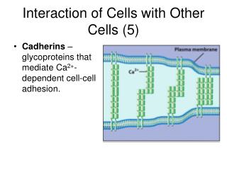
Interaction of Cells with Other Cells (5) • Cadherins –glycoproteins that mediate Ca2+-dependent cell-cell adhesion.
Interaction of Cells with Other Cells (6) • Cadherins (continued) • Also involved in transmitting signals from the ECM to the cytoplasm. • Mediate many of the changes in adhesive contacts during embryonic development by forming epithelial-mesenchymal transition (EMT).
Interaction of Cells with Other Cells (8) • Adherens Junctions and Desmosomes: Anchoring Cells to Other Cells • Adherens junctions – they form “belts” near apical surface called junctional complex. • Cells of an adherens junction held together by calcium-dependent linkages.
Interaction of Cells with Other Cells (9) • Desmosomes – disk-shaped adhesive junctions between cells. • Found in a variety of tissues. • Contain cadherins that link the two cells across a narrow gap. • Cadherins of desmososme shave different domain structures: desmogelins and desmocollins.
Interaction of Cells with Other Cells (10) • The Role of Cell-Adhesion Receptors in Transmembrane Signaling • The transfer of information across the plasma membrane is transmembrane signaling. • Integrins and cadherins can transmit signals from the extracellular environment to the cytoplasm.
Interaction of Cells with Other Cells (11) • The binding of an integrin with its ligand can induce a responses such as changes in growth potential.
The Human Perspective: The Role of Cell Adhesion in Inflammation and Metastasis (1) • Inflammation is a response to infection or injury but can be triggered inappropriately. • Inflammatory response: • Recruitment of leukocytes to site of injury. • Neutrophils attach to P- and E-selectins. • Neutrophils start to “roll” along wall of vessel.
The Human Perspective: The Role of Cell Adhesion in Inflammation and Metastasis (2) • As neutrophils interact with inflamed venule endothelium, platelet activating factor (PAF) is displayed. • PAF sends a signal to increase the binding activity of some integrins. • Activated integrins cause neutrophils to stop rolling and adhere firmly to wall of vessel.
The Human Perspective: The Role of Cell Adhesion in Inflammation and Metastasis (3) • Cancer is the result of abnormal cell proliferation. • The spread of a tumor to other parts of the body is called metastasis. • Metastatic cells have special cell adhesion properties: • Are less adhesive. • Are able to penetrate several barriers. • Are able to invade normal tissues.
The Human Perspective: The Role of Cell Adhesion in Inflammation and Metastasis (4) • During growth and development of a tumor there is loss of E-cadherin leading to less adhesion. • Changes in the numbers and types of cell-adhesion molecules lead to promote metastasis.
7.4 Tight Junctions: Sealing the Extracellular Space (1) • Tight junctions (TJs) – specialized contacts between epithelial cells. • Located at the very apical end of the junctional complex between adjacent cells. • TJs serve as a barrier to the free diffusion of water and solutes from the extracellular compartment. • Some TJs are permeable to specific ions or solutes.
Tight Junctions: Sealing the Extracellular Space (2) • Occludins are proteins found in TJs. • Claudins form the major structural component of TJs, and may account for selective differences in TJ permeability. • TJs form the blood-brain barrier.
7.5 Gap Junctions and Plasmodesmata: Mediating Intercellular Communication (1) • Gap junctions – sites between animal cells for intercellular communication. • Composed entirely of membrane protein connexin. • Connexins are organized into a complex called connexon.
Gap Junctions and Plasmodesmata: Mediating Intercellular Communication (2) • Gap-junction intercellular communication (GJIC) allows the passage of low-weight molecules. • Gap junctions can allow integration of activities of individual cells into a functional unit. • Compatibility differences between connexins either promote or prevent communication between different cells.
Passage of low-molecular-weight solutes through gap junctions
Gap Junctions and Plasmodesmata: Mediating Intercellular Communication (3) • A new type of communication has been discovered – tunneling nanotubes. • It has been observed in cells growing in culture.
Gap Junctions and Plasmodesmata: Mediating Intercellular Communication (4) • Plasmodesmata are cytoplasmic channels passing through cell walls of adjacent plant cells. • Are lined by plasma membrane. • Contain a central structure, the desmotubule. • Serve as sites of cell-cell communication.
7.6 Cell Walls (1) • Cell walls provide plants protection against mechanical abrasion, pathogens, and osmotic stress. • The fibrous component is cellulose. • Cellulose is organized into microfibrils, which provide rigidity to the cell wall.
Cell Walls (2) • The matrix of the cell wall contains hemicelluloses, pectins, and proteins. • Cell walls arise as a cell plate that forms between the plasma membranes of newly formed daughter cells. • The walls of growing cells are primary walls and allow flexibility lacking in the thicker secondary walls of mature cells.