Download

1 / 31
390 likes | 1.01k Vues
Noninvasive Monitoring in The Intensive Care Unit. Iskander Al-Githmi, MD,FRCSC, FCCP Assistant Professor of Surgery King Abdulaziz University. Learning Objectives. Know the different noninvasive monitoring techniques commonly use in ICU.

E N D
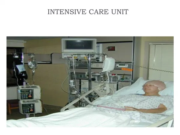
Noninvasive Monitoring in The Intensive Care Unit Iskander Al-Githmi, MD,FRCSC, FCCP Assistant Professor of Surgery King Abdulaziz University
Learning Objectives • Know the different noninvasive monitoring techniques commonly use in ICU. • Know the advantages and limitation of the different noninvasive monitoring methods. • Know the different technologies used in noninvasive monitoring. • Correlate findings observed during noninvasive monitoring with the patient’s changing physiology.
Goals of Monitoring • Assessment of vital organs function • Detection of early life-threatening complications • Determine the needs for intervention.
Respiratory Monitoring • Pulse Oximetry • Capnography
Pulse Oximetry • Measures four types of hemoglobin: deoxyHb, oxyHb, carboxyHb and metHb. • Estimate functional hemoglobin saturation.
Principles of Pulse Oximetry con’t • Spectorphotometry • Discriminate between oxyHb and deoxyHb by the difference in light absorption at 660nm and 940nm • Estimate heart rate by measuring cyclic changes in light transmission.
Pitfalls and Limitations • Margin of error is +/- 4% at Sao2 95% • Margin of error is upto 15% at SaO2 <70% • Does not measure arterial oxygen (PaO2) • Wide Pao2 level 60-160mmHg at O2-Dissociation curve • Is not a substitute for arterial blood gas • SPO2= O2Hb+COHb • SPO2>90%even with COHb 70% • Methylene blue underestimate the saturation • Low perfusion e.g. low cardiac output • Extreme anemia
Capnometry • Clinical practice: • Confirmation of endotracheal tube placement • Estimation of arterial CO2 with End-Tidal CO2 • Monitoring the integrity of patient-ventilator system • Provide a noninvasive means of facilitating weaning from mechanical ventilation
The principles of capnometry • Main stream and side stream analyzer • Infrared spectrometry • CO2 absorption takes place at 4.3 micm
Normal capnogram • Phase I: inspiratory baseline • Phase II: expiratory upstroke • Phase III: expiratory plateau
PaCo2 – PETCo2 gradient • PETCo2 is usually 1-3mmHg lower than PaCo2 • The difference between PETCo2 and PaCo2 is caused by V/Q mismatch • PETCo2 does not reflect PaCo2 in the presence of V/Q mismatch • PaCo2 – PETCo2 gradient is usually < 5mmHg • The gradient increased when cardiac output decreased
Limitations • Alteration of dead space ventilation • Breathing patterns • Patient stability • Tidal volume • V/Q ratio
Sudden loss ETCO2 Possible causes: • Airway disconnection • Dislodgment of ET tube • Total obstruction of ET tube
Gradual decrease in ETCO2 to non zero level Possible causes: • Leak in the circuit • Partial disconnection from ventilator
Gradual decrease in ET CO2 Possible causes: • Hypothermia • Pulmonary embolism • Cardiopulmonary bypass
Rise in baseline ETCO2 Possible causes: • Defective exhalation valve • Rebreathing of previously exhaled CO2
Question 1 The capnograph also has a use in correctly and exactly identifying end expiration during the analysis of haemodynamic wave forms. This would classically be seen when measuring the Pulmonary Artery Wedge Pressure at end expiration.The point of end expiration is seen at: • Just after the peak on the capnography waveform. • Just before the peak on the capnograph waveform. • In the exact middle of the capnography waveform. • In the middle of the capnography "trough".
Question 2 Capnography has a place in confirming endotracheal tube placement but should be used in conjunction with other, simpler techniques. What is the most correct sequence of events in confirming endotracheal tube placement? • Visualise the tube with laryngoscopy / observe and auscultate the chest and epigastrium / check the capnogram. • Visualise the tube with laryngoscopy / check the capnogram / observe and auscultate the chest and epigastrium
Question 3 The Capnograph may also be used in the ICU to avoid constant arterial sampling for ABGs and also to monitor patients with brains injuries who need to be kept with a normal PaCO2.This is possible because of a correlation between the maximum partial pressure of CO2 (PetCO2) at end expiration and the arterial CO2 levels. With normal ventilation and perfusion the gradient between PetCO2 and PaCO2 should be between 1-5 mm Hg.The following statement is most true: • Capnography is unreliable in the case of massive pulmonary embolism. • Capnography is unreliable in the case of ARDS. • Once a PetCO2 to PaCO2 gradient is established, capnography completely obviates the need for ABGs. • Once a PetCO2 to PaCO2 gradient is established capnography can be reliable used.
Question 4 Cardiac arrest can produce a flat capnograph trace, due to low pulmonary blood flow, even if the endotracheal tube is correctly placed above the carina. • True • False
Question 5 Severe bronchospasm can produce a flat capnograph trace even if the endotracheal tube is correctly placed. • True • False
Question 6 12 year-old was rescued by a firefighter form a smoke field room of a burning 12-story apartment building. The following procedures are appropriate, except which of the following: • a careful inspection of the upper airway passage to detect signs of inhalation injury • A chest x-ray on admission to look for inhalation injury • A 24 hour hospital observation • A normal oxygen hemoglobin saturation by pulse oximetry to exclude the presence of CO poisoning
Question 7 The following statement about pulse oximetry are true except which of the following: • Modern pulse oximetry uses two wavelength of light, red and infrared to discriminate between oxygenated and deoxygenated blood • Pulse oximetry is not affected by low cardiac output state • Different form of dyshemoglobinemia can affect the accuracy of oxyhemoglobin measurement by pulse oximetry • The pulse oximetry degrades with oxygen saturation <65%