Download
1 / 45
530 likes | 1.13k Vues
INTENSIVE CARE UNIT. Isaac Asimov: “Life is pleasant. Death is peaceful. It is the transition that is difficult”. Isa a c Asim ov: Prof e ssor o f Bi o ch e mistry B o ston.

E N D
IsaacAsimov:“Life is pleasant. Deathispeaceful.Itisthetransitionthat is difficult” IsaacAsimov:ProfessorofBiochemistryBoston
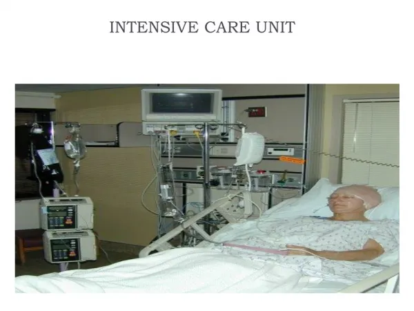
Definition: Is the branch of Medicine concerned with the management of life threatening conditions requiring monitoring of vital signs and usage of sophisticated equipments to keep the patient alive. Intensive Care is usually offered for potentially reversible condition. This is done in designated units called The Intensive Care Units or The Intensive Therapy Units or Critical Care Units. It is costly, because it contains the most expensive, technologically advanced equipments.
. Problems that might need critical care treatment include *cardiac (myocardial infarction , shock, cardiac arrhythmias, congestive heart failure , high blood pressure, and unstable angina). *nervous (e.g. C.V.A) *pulmonary ( acute respiratory failure, pulmonary emboli, hemoptysis ), *endocrine (hormonal) systems (e.g. pheochromocytoma) *complications from surgery *Polytrauma *Severe burns *infections( sepsis,meningitis) *medication monitoring for drug ingestion or overdose .
Monitors, intravenous (IV) tubes, feeding tubes, catheters , ventilators and other equipment are common in critical care units. These can sustain life but can also increase the risk of infection . While patients may recover, death is a possibility for people in critical care.
a. Organization: One has to think carefully about the possibility of establishing an Intensive Care Unit in a hospital, whether the hospital is fit to establish such a unit and whether there are the all the Specialties needed and whether they are prepared to answer consultations quickly.The ratio is set as 2-10% of the beds of the hospital. ICU should be not more than 14 beds (otherwise, a new another ICU should be established) and not less than 4 bedsThe patient either remains as patient of the referring unit or is regarded as ICU patient.It can be of OPEN PLAN or INDEVIDUAL ROOMS.It should contain designated spaces for store, pharmacy, theatre, isolation, doctor room, nurses room, and whatever spaces it will be found necessary. b- LocationAn ICU can be established anywhere in the hospital, but is preferred to be close to the Emergency Department and or the theatre.
c- StaffingIn the past (still in a lot of hospitals) the Anaesthetist is the best one who suited for running the Intensive Care Units for three reasons. (1) The ICU is regarded as continuation to the theatre work, which the Anaesthetist is doing (2) the Anaesthetist has no interest in bed allocation in the hospital (3) the Anaesthetist knows how to ventilate a patient and to keep him alive during ventilation.Regarding the nurses training, which are the backbone of any ICU, need special training in every aspect of IC work especially in the basic nursing. The ratio is one nurse per patient on the ventilator, one nurse per two patients on monitors only.
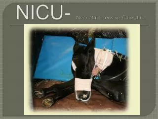
d- TypesApart from the general ICU there are several types of ICU according to the needs of the hospital and availability of staff:1- Neonatal and premature born (NICU)2- Coronary Care Unit (CCU)3- Medical (MICU)4- Neurological (Neuro ICU)5- Trauma (Trauma ICU)6- Post-Anaesthesia (PACU)7- Post-Operative (POCCU) 8- Surgical (SICU)
2) Clinical problems a- Sedation1- I.V. Induction agents (Propofol), disadvantages: bradycardia, reduced systemic vascular resistance. 2- Benzodiazepines (Medazolam, Lorazepam, Diazepam), disadvantages: dependence, withdrawal agitation. 3- Opoids (Morphine, Fentanyl, Alfemtanil, Remifentanil), disadvantages: respiratory depression, bradycardia, hypotension, nausea, constipation. 4- Alpha Agonists (Clonidine), disadvantages: rebound hypertension , bradycardia. b- Pain relief 1- Opioids 2- Epidurals if appropriate
c- Ventilation1- Noninvasive Ventilation2- Invasive Ventilation3- Permissive hypercarbia4- Weaning from mechanical ventilation d- Infection1- The infection the patient brings with him to the ICU2- Ventilator Associated Pneumonia3- Catheter Associated UTI4- I.V.Catheter Associated infection 5- 70% are due to antibiotic resistant organisms
e- DVT and other Thromboemboli1- Early mobilization2- Give low Molecular Weight Heparin (LMWH)3- Stocking , repeated (mechanical) compression of legs f- Ulceration of skin1- Change of position2- Padding of pressure sites h- Stress Ulcers1- Enteral feeding2- Antacids3- Sucralfate4- H2-receptor blockers5- Proton pump inhibitors g- Nutrition : Enteral Parenteral
. Problems that might need critical care treatment include *cardiac (myocardial infarction , shock, cardiac arrhythmias, congestive heart failure , high blood pressure, and unstable angina). *nervous (e.g. C.V.A) *pulmonary ( acute respiratory failure, pulmonary emboli, hemoptysis ), *endocrine (hormonal) systems (e.g. pheochromocytoma) *complications from surgery *Polytrauma *Severe burns *infections( sepsis,meningitis) *medication monitoring for drug ingestion or overdose .
LevelO:normalwardcare • Level1:atriskofdeteriorating,support • fromcriticalcareteam • Level2:moreobservationorintervention,singlefailingorganorpostoperativecare • Level3;advancedrespiratorysupportorbasicrespiratorysupport,multiorgan failure
Although the criteria for admission to an ICU are somewhat controversial—excluding patients who are either too well or too sick to benefit from intensive care—there are four recommended priorities that intensivists (specialists in critical care medicine) use to decide this question. These priorities include: 1- Critically ill patients in a medically unstable state who require an intensive level of care (monitoring and treatment). 2- Patients requiring intensive monitoring who may also require emergency interventions. 3- Patients who are medically unstable or critically ill and who do not have much chance for recovery due to the severity of their illness or traumatic injury. 4- Patients who are generally not eligible for ICU admission because they are not expected to survive. Patients in this fourth category require the approval of the director of the ICU program before admission. WHICH SYSTEM FAILURE IS COMMON TO ALL PATIENTS IN I.C.U.? Though a wide range of pathological conditions required intensive therapy but almost all involve failure of respiratory &/or circulation.Therefore,the treatment of acute respiratory insufficiency represent a major area in ICU. T • Who should be admitted to I.C.U.?
Respiratory indication for admission to ICU 1- Damage of inspiratory center :e.g,drug intoxication , status epilepticus , eclampsia. 2-Upper motor neuron lesion : high cervical spinal lesion. 3- Lower motor neuron lesion : poliomylitis. 4- Peripheral nerves diseases :Guillian Barre syndrome , Tetanus. 5- Neuromuscular junction diseases : Myasthenia gravis. 6- Stractural impairment of chest wall : Flial chest. 7- Upper respiratory tract lesion :epiglotitis , vocal cord paralysis , croup ( laryngotracheobronchitis ). 8- Lower respiratory tract disease : status asthmaticus , acute on chronic lung disease (COPD) , aspiration ( gastric content ,mecunium , foreign body ). 9- Aleveolar disorders (paranchymal dis.) :pulm.oedema , near drowning , ARDS.
Mechanical VentilationMechanical ventilation is a life support treatment. A mechanicalventilator is a machine that helps people breathe when they are notable to breathe enough on their own. Why are ventilators used? To get oxygen into the lungs and body and to help the lungs get rid of carbon dioxide by either: 1.To ease the work of breathing—Some people can breathe but it is very hard. They feel short of breath and Uncomfortable (assisted ventilation). 2. To breathe for a patient who is not breathing ( control mechanical ventilation) How are patients on ventilators monitored? Anyone on a ventilator in an ICU setting will be hooked up to a monitor that measures heart rate, respiratory rate, blood pressure, and oxygen saturation. Other tests that may be done include chest-x-rays and blood drawn to measure oxygen and carbon dioxide (“blood gases”). How long is a ventilator used? A ventilator can be life saving, but its use also has risks. It also doesn’t fix the primary disease or injury; it just helps support a patient until other treatments become effective. patients get off the ventilator at the earliest possible time. “Weaning” refers to the process of getting the patient off the ventilator. Some patients may be on a ventilator for only a few hours or days, while others may require the ventilator for longer. Some patients never improve enough to be taken off the ventilator completely.
Oxygen is widely available and commonly prescribed by medical and paramedical staff. When administered correctly it may be life saving, but oxygen is often given without careful evaluation of its potential benefits and side effects. Like any drug there are clear indications for treatment with oxygen and appropriate methods of delivery. Inappropriate dose and failure to monitor treatment can have serious consequences . . To ensure safe and effective treatment prescriptions should cover theflow rate, delivery system, duration, and monitoring of treatment.Tissues require oxygen for survival. Delivery depends on adequate ventilation, gas exchange, and circulatory distribution. Tissue hypoxia occurs within 4 minutesof failure of any of these systems because the oxygen reserves in tissue and lung are relatively small .
1.Cardiac and respiratory arrest2. Hypoxaemia (Pao2<7.8 kPa, Sao2<90%) 3. Hypotension. 4. Low cardiac output and metabolic acidosis (bicarbonate<18mmol/l) 5. Respiratory distress (respiratory rate >24/min) Recommendations for instituting oxygen therapy
In many acute conditions (for example, asthma, pulmonary embolus), inspired oxygen concentrations of 60-100%for short periods may preserve life until more specific treatment can be instituted. Thereafter oxygen should be given at a dose that will correct hypoxaemia and minimize side effects (increase the Pao2 to 8.0-10.6 kPa). When necessary, oxygen must be given continuously.High dose oxygen given to patients with chronic obstructive pulmonary diseasewho have type II respiratory failure can reduce the hypoxic drive to breathe and increase ventilation-perfusion mismatching. This causes carbon dioxide retention and a respiratory acidosis that may be lethal. In these patients initial treatment with low oxygen concentrations(24-28%)should be progressively increased on the basis of repeated blood gas analysis with the aim of correcting hypoxaemia to a Pao2>6.65 kPa without decreasing arterial pH below 7.26. Non-invasive positive pressure ventilation and respiratory stimulants may help achieve adequate oxygenation and prevent carbon dioxide retention by raising minute ventilation in patients with type II respiratory failure .
Oxygen masks simple facemask( Low flow masks ): is the most commonly available oxygen mask to the public. This has a number of vents on both sides and can deliver 35-40 percent of oxygen. However if the oxygen flow increases to 10L/min, this can deliver up to 50 percent oxygen. The disadvantage with this type of oxygen mask is that this seals poorly and has large ventilation holes, thus oxygen flow is diluted with air. . This oxygen mask is used for non-life threatening conditions
The venture mask ( High flow, jet mixing masks,) , is an oxygen mask that uses a mechanical venturi effect in order to increase the flow rate of oxygen into the mask. . These masks are useful for accurately delivering low concentrations of oxygen (24-35%). When oxygen passes through a narrow orifice it produces a high velocity stream that draws a constant proportion of room air through the base of the venturi valve. Air entrainment depends on the velocity of the jet (the size of orifice and oxygen flow rate) and the size of the valve ports. They provide the total ventilatory requirement unaffected by the pattern of ventilation . In patients with chronic obstructive pulmonary disease and type II respiratory failure these masks reduce the risk of carbon dioxide retention while improving hypoxaemia. They are loose fitting and comfortable to wear. Rebreathing of expired gas is not a problem because the mask is flushed by the high flow rates
Nasal prongsare simple and convenient to use. The Fio2 depends on the flow rate of oxygen (1-6 l/min) and varies according to ventilatory minute volume. At an oxygen flow rate of 2 l/min the oxygen concentration in the hypopharynx of a resting subject is 25-30%. Nasal prongs prevent rebreathing, are comfortable for long periods, and allow oxygen to be continued during talking and eating. Local irritation and dermatitis may occur with high flow rates.
Non-invasive assisted ventilation—Supplemental oxygen may be provided through tight fitting nasal or full face masks during nasal intermittent positive pressure ventilation and continuous positive airways pressure. These techniques have been used to support ventilation in sleep associated hypoventilation, during weaning from mechanical ventilation, and in respiratory failure associated with chronic obstructive pulmonary disease.
.Humidification of oxygenWhen oxygen is delivered at a flow rate of 1-4l/min by mask or nasal prongs, the oropharynx or nasopharynx provides adequate humidification. At higher flow rates or when oxygen is delivered directly to the trachea humidification is necessary. Monitoring oxygen treatmentOxygen treatment can be monitored by blood gas measurements or non-invasively by pulse oximetry. Blood gas analysis provides accurate information on the pH, Pao2, and Paco2. Oximetry provides continuous monitoring of the state of oxygenation.Oxygen should be stopped when :1- arterial oxygenation is adequate with the patient breathing room air (Pao2>8 kPa, Sao2>90%).2- In patients without arterial hypoxaemia but at risk of tissue hypoxia, oxygen should be stopped when the acid-base state and clinical assessment of vital organ function are consistent with resolution of tissue hypoxia.
Dangers of oxygen treatmentFire: Oxygen promotes combustion. Facial burns and deaths of patients who smoke when using oxygen are well documentedPulmonary oxygen toxicity: High concentrations of oxygen (>60%) may damage the alveolar membrane when inhaled for more than 48 hours. Progression to the adult respiratory distress syndrome with high protein alveolar oedema and pulmonary radiographic infiltrates is associated with high mortality
Respiratory Failure Defined as : inability of the lung to maintain arterial oxygen tension with or withoutacceptable elimination of CO2 adequets for the patients metabolic requirement Diagnosis : 1/ Clinically A-The disease itself may direct the attention for the diagnosis,e.g.Guillian Barre syndrom,myasthenia gravis , etc. B-The effect of hypoxaemia on CNS , CVS ,RS.: On CNS:ranging from restlessness to coma . On CVS:dusky colouration of the skin and mucous memb,cyanosis or pallor(due to overactivity of sympathetic system). On Respiratory system:Rapid shallow or irregular and grunting Note:- restlessness may be the only obvious sign of hypoxaemia,and if such patient is misdiagnosed and restlessness is treated by sedative drugs, this may leads to rapidly progressive respiratory failure. 2/Blood Gas Analysis :-low PaO2 with ( low ,normal or increased PaCO2 according to the type of respiratory failure) • 3/pulm.function test : decrease in vital capacity , maximum inspiratory force . FEV1 TYPES OF RESP.FAILURE: Type 1 :low PaO2 with low or normal PaCO2. Type 2 :low PaO2 with high PaCO2.
TREATMENT OF ACUTE RESPIRATORY FAILURE: 1- Supplemental oxygen (maintain PaO2 60 – 80 mmHg). 2-Intubation of the trachea. 3-mechanical support of ventilation( adjust the ventilator setting according to arterial blood gas analysis i.e,PaO2,PaCO2 and PH ). 4- Diuretics: in some cases. 5- Optimize intravascular fluid volume (guidelines include CVP , bl.pr. , Urine output and body weight). 6- Inotropic support of cardiac function ( to offsets the adverse of IPPV on CVS ). 7- Removal of secretions 8- Control of infection. 9-Nutritional support . 10- Prophylactic antiacids and /or H2 antagonist
Total parenteral nutrition (TPN) A. Aseptic placement of central venous catheter prior to initiation of TPN. B. Do not add drugs to TPN. If necessary use multi line system. C. Use bacterial filter to prevent infection. D. Calculate ideal body weight/height: Male: 50 + 2.3 kg for each inch in length over 5 feet (60 inches) Female: 45 + 2.3 kg for each inch in length over 5 feet (60 inches) If the actual body weight is greater than 30% of the calculated IBW, calculate the adjusted body weight (ABW): ABW = IBW + 0.4(actual weight - IBW)
E. Nutritional calculation: (per day) When designing a TPN solution, you need to consider these questions: 1. What is the patient’s energy need? (Kcal required/day) 2. How much protein/nitrogen does the patient need in a day? 3. How much fluid can the patient tolerate and need? 4. How much fat emulsion can be given/tolerated? 5. How much dextrose is needed? Dextrose concentration? 6. Which electrolytes are needed and how much? Which vitamins and mineral are needed and how much? What is the osmolality of the solution? What is the route of feeding used?
1. DETERMINING ENERGY NEEDS: Normal need: 25-30 Kcal/kg/day Elective surgery: 28-30 kcal/kg/day Severe injury: 30-40 kcal/kg/day Extensive trauma/burn: 45-55 kcal/kg/day Example: for a 70 Kg male with elective surgery X 30 Kcal/Kg = 2,100 Kcal/day 2. DETERMINE THE PROTEIN NEEDS: 1.5 g/kg/day ( or nitrogen 0.1-0.2 mg/kg/day according to catabolic state i.e. degree of burn, sepsis or trauma). Another way to estimate protein needs is to use the non-protein energy (Kcal) to nitrogen (N) ratio. The non-protein energy to N ratio is based on the premise that sufficient energy must be ingested before protein will be used for tissue maintenance and repair. The ratio of 100-150 Kcal: 1 gm N in stressful condition promote anabolism and 250-300 Kcal: 1 gm N is adequate for normal body maintenance. • If your 70 Kg patient needs 2100 Kcal and you want an Energy:N ratio of 150:1, how many grams of N are you going to need? 2100 Kcal ÷ 150 Kcal/gm N = 14gm N • NOTE: 6.5 gm protein contains 1 gm N. • In our examples: 14 gm N × 6.5 = 87.5 gm protein. • How much protein in the solution? Assume that we give this 70 kg person 85 gm of protein using 10% amino acid solution:= 850 ml
“VAMIN 18 g N/l electrolyte free infusion fluid ”: contain 114 g protein/l = 18 g N/l = 460 kcal). Up to 1000 ml i.v. per day depending on patient’s requirement. Infusion time for 1000 ml Vamin should be at least 8 hrs (maximum infusion rate 2.1 ml/min). 1 gm protein provides 4 kcal.
Contrindications: • Congenital disturbance of amino acid metabolism • Irreversible liver damage • Serious uraemia when there is no possibility of dialysis Precaution: Vamin 18 g N/l electrolyte-free has a high osmolality and should thus not be administered undiluted in peripheral veins. In the event of peripheral administration, the risk of thrombophlebitis can be reduced by means of simultaneous administration of intralipid. Overdose: Nausea and a feeling of heat may occur in the event of infusion of amino acids. especially if the solution is infused too quickly. The symptoms usually disappear if the speed of infusion is lowered.
3- CALCULATE THE AMOUNT OF FLUIDS THE PATIENT NEEDS: either by a/100 cc free water/gram N intake + 1 cc/Kcal provided. E.g. 14 g N X 100 = 1,400 cc + 2100 cc for each Kcal provided =3600 cc/day. b/minimum of 30 cc fluid/Kg body weight to maintain hydration and 50 cc fluid /kg body weight for good hydration. E.g. 50X 70= 3500 cc/day. 4. THE AMOUNT OF FAT NEEDED DEPENDS ON THE TOTAL ENERGY NEED: 1.0 to 2.5 gram fat/Kg. MAXIMAL tolerance level of lipid is considered to be 2.5 gm/Kg body weightand 60% of energy from fat is also considered to be upper limit. More than 60% of energy from fat may result in hyperlipidemia due to impaired lipid clearance by the body. Fat has 9 Kcal/gram. 10% fat emulsion has 1.1 Kcal/cc 20% fat emulsion has 2.0 Kcal/cc 30% fat emulsion has 3.0 Kcal/cc Fat emulsions are available most commonly in 250cc and 500 cc bottle.
Example: What is the energy content of a 500 cc bottle of 10% fat emulsion? 500cc X 1.1 Kcal/cc = 550 Kcal Energy content of a 500 cc of 20% fat? 500 cc X 2.0 Kcal/cc = 1000 Kcal Fat has 9 Kcal/gram so you can determine the number of fat grams if you need it. e.g. How many grams of fat in 500 cc of 10% lipid fat? 500 cc X 1.1 Kcal/cc = 550 Kcal; 500 Kcal × 9 Kcal/gram = 61 gram of fat. 5- CARBOHYDRATE IS THE MAIN SOURCE OF FUEL TO MEET ENERGY NEEDS. Minimal amount of CHO needed per day is 100 grams. dextrose monohydrate yields 3.4 Kcal/gram Glucose yields4 Kcal/gram The amount of fat used in solution will determine the amount of CHO needed. Energy content of one liter (1000 cc) 50% Dextrose solution (D50W)? 1,000 cc X 50% = 500 gram dextrose X 3.4 Kcal/gram = 1,700 Kcal from CHO Energy content of 1000 cc of D5W (5% dextrose solution)? 1,000 cc X 0.05 = 50 gm dextrose X 3.4 Kcal/gram = 170 Kcal
6. Monitoring: • C-reactive protein, Magnesium, Ph, prealbumine, triglyceride level. b. Signs and symptoms of acute lipid intolerance : fever, chills, vomiting, urticarial, chest and back pain. c. Signs and symptoms of rapid infusion reaction to lipid: palpitation, tachypnea, wheezing, cyanosis, nausea, pain at the injection site, headache and oily taste in mouth. d. Signs and symptoms of fluid overload or dehydration 7. Gradual weaning from TPN to enteral or oral route.
Intralipids (fat emulsions) may be infused with TPN simultaneously, if not already admixed with TPN. The primed lipid tubing shall be attached to the y-connection at the catheter hub. When lipids are infused in this manner, the lipid infusion shall be completed within 12 hours of initiation. Exception: For Pediatrics, lipids shall be completed within 20 hours of initiation. Infusion Pump and Filters TPN solutions shall be infused via an infusion control device utilizing the appropriate filter. Prepared mixtures of TPN and Lipids shall be infused with a 1.2 micron, 24 hour filter, and TPN solutions which are not mixed with lipids shall be infused with a .2 micron, 72 hour filter. When the patient is alert, instructs patient to perform Valsalvamaneuverwhile replacing tubing (To enhance positive pressure of the central vessels, thereby reducing risk of air embolism).