Intensive Care Unit Introduction
660 likes | 1.9k Vues
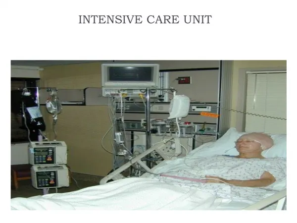
Intensive Care Unit Introduction. Dr Amr Kandeel Director General, Communicable Disease Control Department Director, National Infection Control Program. ICU patients. Sickest patients (multiple diagnoses, multi-organ failure, immunocompromised, septic and trauma) Move less Malnourished

Intensive Care Unit Introduction
E N D
Presentation Transcript
Intensive Care UnitIntroduction Dr Amr Kandeel Director General, Communicable Disease Control Department Director, National Infection Control Program
ICU patients • Sickest patients (multiple diagnoses, multi-organ failure, immunocompromised, septic and trauma) • Move less • Malnourished • More obtunded (Glasgow coma scale) • Diabetics and Heart failure
ICU Care is Invasive • More invasive lines and procedures including surgeries • Longer length of stay • More IV and parenteral drugs • More tube feeding and Parenteral nutrition • More ventilation
ICU : Factors that increase cross-infections • Hand washing facilities • Patient close together or sharing rooms • Understaffing • Preparation of IVs on the unit • Lack of isolation facilities • No separation of clean and dirty AREAS • Excessive antibiotic use • Inadequate decontamination of items & equipments • Inadequate cleaning of environment
Some Health-Care Associated Infections that May Occur • UTI associated with Foley catheters • Lower respiratory tract infection (post-op and ventilator dependent) • Skin necrosis (skin breakdown) • Blood stream infection (and line associated) • Surgical-site infection • Nutrition-related and malnutrition
Strategy for Prevention • Handwashing • Use gloves to prevent contamination of the hands when handling respiratory secretions • Wear gloves and gowns (contact precautions) during all contact with patients and fomites potentially contaminated with respiratory secretions • Use aseptic technique
Strategy for Prevention • Clean and decontaminate all equipment after use • Sterilise or use high-level disinfection for all items that come into direct or indirect contact with mucous membranes • Rinse and dry items that have been chemically disinfected • Package and store items to prevent contamination before use • Keep environment clean, dry and dust free
Conclusions : Strategy for Infection Prevention • Strict attention to Hand hygiene • Prudent Antibiotic use • Aseptic technique • Disinfection/Sterilization of items and equipment • Education of staff infection control awareness • Keep Environment Clean, Dry and dust free • Surveillance of nosocomial infection to identify problems areas & set priorities
Central Venous Catheters Indications • IV fluids and drugs • Blood and blood products • Total Parenteral Nutrition (TPN) • Haemodialysis • Haemodynamic monitoring
Serious Infective Complications • Blood Stream Infections (BSI) • Septic pulmonary emboli • Metastasis infection • Acute endocarditis • Osteomyelitis • Septic arthritis • Shock and organ failure • Poor outcome: Staph.aureus or Candida spp.
Incidence of CR-BSI • Type of catheter Teflon or Polyurethane ( < infections) vs Polyvinyl chloride or Polyethylene • Site of insertion Subclavian (< infections) vs Internal Jugular & Femoral (high risk of colonization & deep venous thrombosis) • No. of Lumen Single-lumen catheter (< infections) vs Multi-lumen catheter
Intrinsic contamination of infusion fluid Port for additives Connection with administration set Insertion site Injection ports Administration set connection with IV catheter Sources of Infection
Sources of Infection 1. Extraluminal Spread • Patient’s own skin micro flora • Microorganism transferred by the hands of Health Care Worker • Contaminated entry port, catheter tip prior or during insertion • Contaminated disinfectant solutions • Invading wound 2. Intraluminal Spread Intralumunal Spread • Contaminated infusate (fluid, medication) • Contaminated infusate (fluid, medication) Skin attachment Skin Fibrin Vein 3. Haematogenous Spread • Infection from distant focus
Prevention of CR-BSI Written Protocol Must be performed by trained staff according to written guidelines Sterile procedure Sterile gown, Sterile gloves, Sterile large drapes Don't shave the site Hand disinfection With an antiseptic solution eg Chlorhlexidine gluconate
Prevention of CR-BSI Skin antisepsis • 2% Chlorhlexidine gluconate has shown to have lower BSI than 10% Povidone-iodine or 70 % Alcohol • 2-min drying time before insertion Maki DG et al. Lancet 1991;338:339-43 • No difference between 0.5% Chlorhlexidine gluconate or 10% Povidone-iodine Humar A et al. Clin Infect Dis 2000;31:1001-7
Prevention of CR-BSI Dressing • Gauze dressings every 2 days • Transparent dressing every 7 days on short term catheter • Replace dressing when catheter is replaced or dressing becomes damp or loose. Grady NP et al, HICPAC draft guidelines: 2002
Prevention of CR-BSI Catheters removal • Don’t replace it routinely • Replace it if: • Inserted in an Emergency • Non functioning • Evidence of local or systemic infection General handling • Opening of hub: Use antiseptic-impregnated pads eg Chlorhexidine gluconate or povidone iodine
Prevention of CR-BSI Administration sets • Replacement at 72-h intervals • No difference in phlebitis if left for 96 hours • Lines for lipid emulsion: replacement at 24-h intervals • Lines for blood product : remove immediately after use
Prevention of CR-BSI Topical antibiotic • Prophylactic use of topical Mupirocin (Bactroban) at insertion site or in nose is not recommended • Rapid development of Mupirocin resistant • Mupirocin affect the integrity of Polyurethane catheter Systemic antibiotic • Prophylactic use of antibiotic is not recommended at the time of catheter insertion
External urethral meatus & urethra • Pass catheter when bladder full for wash-out effect. • Before catheterization prepare urinary meatus with an antiseptic ( e.g. povidone iodine or 0.2% chlorhexidine aqueous solution) • Inject single-use sterile lubricant gel (e.g. 1-2%) lignocaine into urethra and hold there for 3 minutes before inserting catheter. • Use sterile catheter. • Use non-touch technique for insertion
Junction between catheter & drainage tube • Do not disconnect catheter unless absolutely necessary. • For urine specimen collection disinfect outside of catheter proximal to junction with drainage tube by applying alcoholic impregnated wipe and allow it to dry completely then aspirate urine with a sterile needle and syringe.
Junction between drainage tube & collection bag • Keep bag below level of bladder. If it is necessary to raise collection bag above bladder level for a short period, drainage tube must be clamped temporarily. • Empty bag every 8 hours or earlier if full. • Do not hold bag upside down when emptying
Tap at bottom of collection bag • Collection bag must never touch floor. • Always wash or disinfect hands (eg with 70% alcohol) before and after opening tap. • Use a separate disinfected jug to collect urine from each bag. • Don't put disinfectant into urinary bag.
Incidence of HAI vs. Cost Haley, 1986
Risk factors for bacterial pneumonia Host FactorsFactors that facilitate reflux & aspiration into the lower RT • Elderly • Severe Illness • Underlying Lung Disease - Mechanical ventilation • Depressed Mental Status - Tracheostomy • Immunocompromising - Use of a Nasogastric Tube Conditions or Treatments - Supine Position • Viral Respiratory Tract Factors that impede normal Infection Pulmonary Toilet Colonisation- Abdominal or thoracic surgery • Intensive Care Setting - Immobilisation • Use of Antimicrobial Agents • Contaminated hands • Contaminated Equipment
Prevention in ICU • Turn patients to encourage postural drainage • Encourage to take deep breaths and cough. • Maintain an upright position (elevate patient’s head to 30º- 45º degree angle) to reduce reflux and aspiration of gastric bacteria.
Gastric Ulcer Prophylaxis • Stomach of a healthy person : Acidic pH () & normal peristalsis movement prevent bacterial growth • Alkaline pH () and loss on normal peristalsis lead to bacterial colonisation which increases the risk of ventilator-associated pneumonia • Mechanical ventilation patients are at increased risk for upper GI haemorrhage from stress ulcers. • H2 blockers or antacids are used to prevent stress ulcers
Nasogastric Tube • May erode the mucosal surface • Block the sinus ducts • Regurgitation of gastric contents leading to aspiration. • Verify placement of the feeding tube in the stomach or small intestine by X ray • Elevate the head of the bed 30º- 45 º degrees Remove NG Tube if not necessary
Ventilators • After every patient, clean and disinfect (high-level) or sterilize re-usable components of the breathing system or the patient circuit according to the manufacturer’s instructions.
Suctioning mechanically ventilated patients • Handwashing before and after the procedure. • Wear clean gloves to prevent cross-contamination • Use a sterile single-use catheter ; if it is not possible then rinse catheter with sterile water and store it in a dry, clean container between uses and change the catheter every 8 - 12 hours.
Suction Bottle • Use single-use disposable, if possible • Non-disposable bottles should be washed with detergent and allowed to dry. Heat disinfect in washing machine or send to Sterile Service Department.
Nebulizers • Use sterile medications and fluids for nebulization • Fill with sterile water only. • Change and reprocess device between patients by using sterilization or a high level disinfection or use single-use disposable item. • Small hand held nebulizers • minimise unnecessary use • between uses for the same patient disinfect, rinse with sterile water, or air dry and store in a clean, dry place • Reprocess nebulizers daily
Humidifiers • Clean and sterilize device between patients. • Fill with sterile water which must be changed every 24 hours or sooner, if necessary. • Single-use disposable humidifiers are available but they are expensive.
Oxygen mask • Change oxygen mask and tubing between patients and more frequently if soiled
Antibiotics use Must avoid widespread use of broad spectrum antibiotics
Examples illustrating difficult to detect infections: Long Incubation period Hard to detect HIV, Hep B or Hep C due to long incubation period Easier to detect Staph aureus food borne illness, or toxic shock due to re-use of medication vial. These infections occur 1 hr to days post event.
Examples of difficult to detect infections: Uncultivable organisms Viruses are under appreciated as causes of nosocomial infections. Except in cases of high morbidity viral cultures are not done in resource scarce settings. Impact food-borne, respiratory, water borne illnesses. We don’t know the spectrum of anti-microbial activity of most preservatives and cleaners for many viruses.
Define the problem to tracked • Advantages: • The occurrence then can be compared in different facilities and in different time periods • Definitions can be suspect, probable or confirmed depending upon the information that is available
Examples from the NNIS Manual • Symptomatic Urinary Tract Infection: • Patient must have one of the two criteria: • Fever >38 C OR urgency OR frequency OR dysuria OR suprapubic tenderness without other cause OR • Urine culture with at least 105 organisms per ml or no more than two species of organisms
Definition of surgical site infection (no implant) • Occurs within 30 days of surgery AND has one of the following: Purulent drainage from drain OR Organism isolated from aseptically obtained fluid in the organ space
Example of lab-confirmed blood stream infection • Patient has a recognized pathogen cultured from one or more blood cultures AND • Organism cultured is not related to another infection at another site
Prior to starting any surveillance • Agree upon a written case definition that is practical given the laboratory facilities and patient work load in your facility.