Download
1 / 91
920 likes | 1.08k Vues
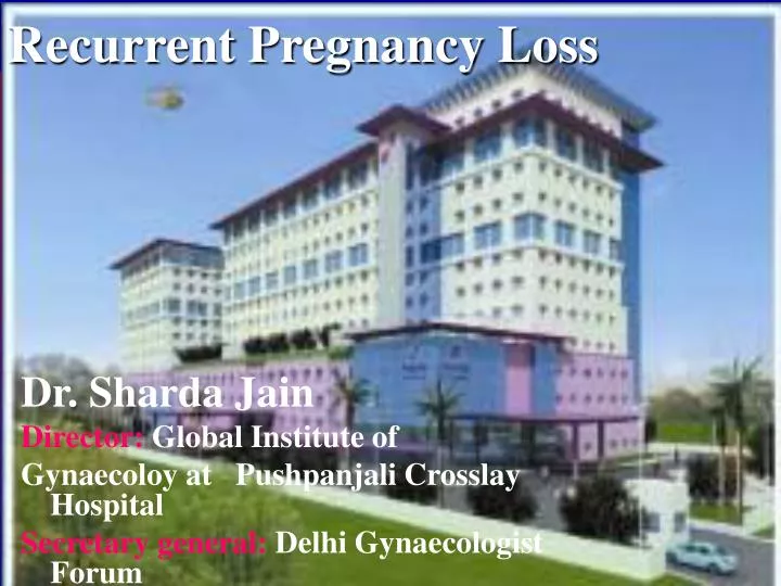
Recurrent Pregnancy Loss. Dr. Sharda Jain Director: Global Institute of Gynaecoloy at Pushpanjali Crosslay Hospital Secretary general: Delhi Gynaecologist Forum. Learning Objectives. Identify possible causes of early pregnancy loss

E N D
Recurrent Pregnancy Loss Dr. Sharda Jain Director:Global Institute of Gynaecoloy at Pushpanjali Crosslay Hospital Secretary general:Delhi Gynaecologist Forum
Learning Objectives • Identify possible causes of early pregnancy loss • Outline basic evaluation for recurrent pregnancy loss (RPL) • Review current treatment approaches for these patients
Definition • Classical: 3 or more consecutive pregnancy losses before 20 weeks gestation • Expanded: 2 or more consecutive losses • Risk of further loss similar for 2 versus 3 consecutive losses • Initiation of evaluation appropriate after 2 losses based on patient age and desire Hill Curr Prob Obstet Gynecol Fertil 1994;37:693-704
Recurrent Loss Epidemiology • 5% of couples attempting pregnancy have 2 or more consecutive losses • 1% have 3 or more consecutive losses • Most clinicians consider RPL even if losses are not consecutive Lee Semin Reprod Med 2000;18(4):433-40
SPAB Epidemiology • 34% pregnancy loss in prospective cohort of healthy women • 22% unrecognized - detected by assay only • 12 % clinically recognized • Obstetrical history predictive • prior success: 4-6 % chance of loss • prior loss: 19-24%chance of loss Wilcox NEJM 1988;319:189-194
SPAB or RPL? • A single SAB, unless a successful pregnancy intervenes, increases the risk for the next pregnancy • Distinction between “sporadic” and “recurrent” loss blurred • Effect of maternal age: SAB risk approaches 50% by age 40 for both aneuploid and euploid losses Cramer Semin Reprod Med 2000;18(4):331-9
Miscarriage Recurrence Risk Warburton D, Fraser FC: Am J Human Genet 16:1, 1964
PCOS & Pregnancy Loss • Pregnancy loss ↑ with PCOS • Franks S, Ann Int Med 93, Jacobs HS BRJOBGYN 93 • GnRH-a ↓ miscarriage in PCOS women • Homburg R, et al: Fertil Steril 59:527, 1993 • RSA patients with ↑ LH, DHEAS or T more likely to miscarry • Tulpalla M, et al: BrJOBGYN 100:348, 1993 • GnRH-a ↓ miscarriages in RSA patients with PCOS compared to clomid (10% vs 55%) Johnson P, et al: BMJ 300:154, 1990
Metformin Reduces Pregnancy Loss in PCOS • Retrospective study of PCOS women who became pregnant • Group 1: metformin during pregnancy (n=101) • Group 2: control (n=31) • Early loss rate 12.9% vs 41.9% (p=0.001) • Prior SPAB: 15.7% vs 58.3% (p=0.005) Jakubowicz DJ, et al: abstract P2-427, Endocrine Society, 2001
Etiology Anatomic Factors • 10-15% recurrent 1st trimester losses have congenital anomaly • Variations of the double uterus the most common • Septate loss rates 25-90% - usually amenable to resection • Bicornuate loss rates 40% - uncertain benefit of surgery Propst & Hill Semin Reprod Med 2000;18(4):341-50
Etiology Anatomic Factors • Unicornuate uteri 50% loss • Uterus didelphys 40% loss • DES exposure - many have abnormal uterine structure • Cervical incompetence • Intrauterine synechiae
Etiology Anatomic Factors • Unclear relationship between uterine leiomyomata and RPL • Large submucosal fibroids distort the cavity or occupy a large subendometrial area • ? Mechanism(s) - mechanical constriction or inadequate placentation resulting from poorly vascularized endometrium
Etiology Infection • No infectious agent has been proven to cause recurrent pregnancy loss • ? Colonization with Ureaplasma urealyticum leading to empiric antibiotics • Certain infections have been associated with spontaneous loss • Toxoplasma gondii, rubella, HSV, CMV, measles, coxsackie Lee Semin Reprod Med 2000;18(4):433-40
Etiology Genetic Factors • Trisomy (50%) • #16 all lethal 1/3 of all trisomies • #21 Down Syndrome usually due to meiotic non-disjunction 80% maternatal • Monsomy X (20%) • 45X Turner Syndrome most common • Triploidy (15%) • 90% from father • Tetraploidy (5%) • Mosaicism (2%)
Etiology Genetic Factors • Parental abnormalities in 3-5% of couples with recurrent loss • Balanced translocation most common • Reciprocal (60%) or Robertsonian (40%) • 25-50% risk of pregnancy loss • May eventually produce normal offspring
Etiology Genetic Factors • Homologous Robertsonian translocation • 1/2500 couples • precludes successful reproduction • Heterozygous may lead to partial monosomy or trisomy; “milder” phenotypical expression Ward Semin Reprod Med 2000;18(4):425-32
Etiology Genetic Factors • Speculation about single gene mutations • Blastocyst formation • Implantation • Morphogenesis of vital organs
Etiology Genetic Factors • Skewed X inactivation • Preferential inactivation(>90%) of one of the X alleles • May be lethal to a male offspring • May result in X-autosome translocations • Trisomy mosaicism in the germline
Etiology Genetic Factors • Advanced maternal age • Impact on risk for pregnancy loss cannot be over-emphasized • Increased rates of maternally-derived trisomies • Probable “natural selection” of better quality oocytes earlier in reproductive life • Oocytes recruited later in life more likely to be abnormal or experience meiotic error
Decline in the Number of Oocytes from Birth to Menopause Lobo, R. A. N Engl J Med 2005;353:64-73
Fertility and Miscarriage Rates as a Function of Maternal Age Heffner, L. J. N Engl J Med 2004;351:1927-1929
EtiologyThrombophilia • Pregnancy is a hypercoagulable state • Women with heritable or acquired thrombophilic disorders have significantly increased risks of pregnancy loss Kutteh Semin Reprod Med 2006;24(1):54-65
EtiologyThrombophilia Venous • Most common inherited: • Heterozygous Factor V Leiden (G1691A) • Factor II-prothrombin mutation (G20210A) • Hyperhomocysteinemia (MTHFR C677T and A1298C)
EtiologyThrombophilia Venous • Most common acquired: • Anti-phospholipid antibodies (APAs) • Activated Protein C resistance • Hyperhomocysteinemia (MTHFR C677T and A1298C) • Other possible abnormalities • Anti-thrombin deficiency • Protein C or S deficiency • Elevated Factor VIII
EtiologyThrombophilia Arterial • Hyperhomocysteinemia • APAs • Lupus anticoagulant
EtiologyThrombophilia • Factor V Leiden • Abnormal factor V resistant to anticoagulant effects of activated protein C • Majority of patients resistant to activated protein C will be heterozygous for Factor V Leiden • Present in 3-8% of the White population • Rare in Blacks, Asians, Native Americans
EtiologyThrombophilia • Factor V Leiden • Autosomal dominant • Acquired activated protein C resistance in pregnancy, OCP use and in presence of APAs • Heterozygotes: 7X increase lifetime risk thrombosis; 15X increase during pregnancy or OCP use • Homozygotes: 50-100X increase lifetime risk thrombosis
EtiologyThrombophilia • Prothrombin G20210A Mutation • Higher plasma prothrombin concentrations, augmented thrombin generation • Heterozygotes: 2-3% Whites • Conflicting prevalence studies among RPL • Recent critical review suggests an association
EtiologyThrombophilia • Hyperhomocysteinemia polymorphisms • C677T thermolabile MTHFR • Heterozygous 10-20% Whites • Normal or slightly elevated homocysteine • Increased homocysteine when combined with B vitamin deficiencies • Homozygous 10% Whites • Significantly increased homocysteine
EtiologyThrombophilia • Hyperhomocysteinemia polymorphisms • A1298C often occurs with thermolabile C677T • 33% frequency in Dutch population • Combined heterozygosity results in hyperhomocysteinemia and decreased plasma folate levels
EtiologyThrombophilia • Hyperhomocysteinemia polymorphisms • Significant association between hyperhomocysteinemia and RPL • ? Mechanism: interference in embryonic development through defective chorionic villous vascularization • Known association with later pregnancy-related complications
EtiologyThrombophilia • Anti-thrombin Deficiency • Physiologic inhibitor of coagulation • Type I: quantitative; decreased antigen and function; caused by gene deletions, nucleotide changes • Type II: qualitative; normal antigen levels, decreased function; caused by point mutations with single amino acid changes leading to a dysfunctional protein
EtiologyThrombophilia • Anti-thrombin Deficiency • Autosomal dominant • Prevalence Type I heterozygous carriers: 1/2000 – 1/5000 • Prevalence Type II heterozygous carriers: 3/1000 • Most thrombogenic of inherited thrombophilia: 20-50% lifetime risk • Associated increased risk stillbirth and fetal loss
EtiologyThrombophilia • Protein C Deficiency • Down-regulates coagulation cascade; deficiencies lead to unregulated fibrin formation • Autosomal dominant: > 160 mutations • Type I: quantitative • Type II: decreased function • Associated with 2nd trimester losses
EtiologyThrombophilia • Protein S Deficiency • Principal cofactor of activated Protein C; mimics C deficiency: questionable association with pregnancy loss • Autosomal dominant: > 160 mutations: prevalence 0.15-0.8% general population; acquired forms in multiple disease states • Type I: quantitative • Type II: decreased function • Type III: low free protein, normal antigen, reduced activity
EtiologyLuteal Phase Defect • Luteal phase defect is a controversial cause of RPL • Studies proving LPD as a cause of RPL lacking • No convincing studies showing LPD treatment improves pregnancy outcome Lee Semin Reprod Med 2000;18(4):433-40 • 80% of women with low midluteal progesterone proceed to term • 20% of fertile women have abnormal endometrial biopsies • P4 drops after meals & standing
Etiology Endocrine Factors • Poorly controlled diabetes • Overt hyperthyroidism • Overt hypothyroidism • No evidence that asymptomatic systemic endocrinologic or metabolic disorders are a cause of RPL
EtiologyAutoimmune Factors • Certain autoimmune diseases are associated with pregnancy loss • Systemic lupus erythematosis • 1st trimester loss: 10% risk • 2nd and 3rd trimester loss: 6% • Anti-phospholipid syndrome • 2nd trimester loss: 38% Fausett & Branch Semin Reprod Med 2000;18(4):379-392
EtiologyAutoimmune Factors • Anti-phospholipid antibodies (aPL) • autoantibodies recognizing various combinations of phospholipids, phospholipid-binding proteins, or both • Anti-phospholipid syndrome (APS) - clinical association between aPL and syndrome of hypercoagulability Levine NEJM 2002;346:752-63
EtiologyAutoimmune Factors • APS diagnostic criteria: • Clinical features • Vascular thrombosis or • Loss of fetus at or after 10 weeks or • Preterm delivery at or before 34 weeks or • 3 or more consecutive SAB before 10 weeks
EtiologyAutoimmune Factors • APS diagnostic criteria: • Laboratory features • Anti-cardiolipin (aCL) antibodies: IgG or IgM at moderate or high levels on 2 or more occasions at least 6 weeks apart • Lupus anticoagulant (LA) antibodies: detected on 2 or more occasions at least 6 weeks apart
EtiologyAutoimmune Factors • Other anti-phospholipid antibodies • Anti-phosphatidylserine: nearly always associated with APS, highly correlated to cardiolipin binding • Other antibodies have less correlation • No consistency among reported studies • No independence from aCL Fausett Semin Reprod Med 2000;18(4):379-92
EtiologyAutoimmune Factors • Low levels of aPL are not associated with RPL • Assays for non-aCL aPL are not standardized • Studies thus far are contradictory
EtiologyAutoimmune Factors • Other auto-antibodies NOT associated with RPL • Anti-nuclear antibodies may be more common among women with RPL but their presence or absence do not predict subsequent pregnancy outcome
EtiologyAutoimmune Factors • Other auto-antibodies NOT associated with RPL • Anti-thyrogobulin and anti-thyroid peroxidase are markers of increased risk for pregnancy loss if identified early in pregnancy • Some small studies suggest a slight association in RPL; other larger studies do not • Subsequent pregnancy outcomes not affected
EtiologyAlloimmune Factors • Immune response to non-self components of pregnancy • Cytotoxic antibodies • Absence of maternal blocking antibodies • Inappropriate sharing of HLA • Disturbances in natural killer cell function and distribution Porter Semin Reprod Med 2000;18(4):393-400
EtiologyAlloimmune Factors • Cytotoxic antibodies • Maternal response to paternal antigens • Present in normal pregnancies • More common in fertile couples than those with RPL • No bearing on subsequent pregnancy outcome